 Movie
Movie Controller


+ Open data

- Basic information
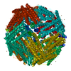
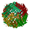
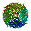
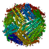
| Entry | Database: PDB / ID: 6s61 | ||||||
|---|---|---|---|---|---|---|---|
| Title | Apoferritin from mouse at 1.84 angstrom resolution | ||||||
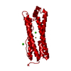
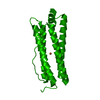
 Components Components | Ferritin heavy chain | ||||||
 Keywords Keywords | METAL BINDING PROTEIN / iron ion binding / ferroxidase / iron storage / cytoplasm | ||||||
| Function / homology |  Function and homology information Function and homology informationIron uptake and transport / Golgi Associated Vesicle Biogenesis / ferroxidase / autolysosome / negative regulation of ferroptosis / ferroxidase activity / negative regulation of fibroblast proliferation / endocytic vesicle lumen / Neutrophil degranulation / ferric iron binding ...Iron uptake and transport / Golgi Associated Vesicle Biogenesis / ferroxidase / autolysosome / negative regulation of ferroptosis / ferroxidase activity / negative regulation of fibroblast proliferation / endocytic vesicle lumen / Neutrophil degranulation / ferric iron binding / autophagosome / iron ion transport / ferrous iron binding / intracellular iron ion homeostasis / immune response / iron ion binding / negative regulation of cell population proliferation / mitochondrion / extracellular region / membrane / identical protein binding / cytosol / cytoplasm Similarity search - Function | ||||||
| Biological species |  | ||||||
| Method | ELECTRON MICROSCOPY / single particle reconstruction / cryo EM / Resolution: 1.84 Å | ||||||
 Authors Authors | Vonck, J. / Pfeil-Gardiner, O. / Mills, D.J. | ||||||
 Citation Citation | Journal: To Be Published Title: To be published later Authors: Vonck, J. / Mills, D.J. / Pfeil-Gardiner, O. | ||||||
| History |
|
- Structure visualization
| Movie |
Movie viewer |
|---|---|
| Structure viewer | Molecule: MolmilJmol/JSmol |
- Downloads & links
-Download
| PDBx/mmCIF format | 6s61.cif.gz | 1.4 MB | Display | PDBx/mmCIF format |
|---|---|---|---|---|
| PDB format | pdb6s61.ent.gz | 1.2 MB | Display | PDB format |
| PDBx/mmJSON format | 6s61.json.gz | Tree view | PDBx/mmJSON format | |
| Others |  Other downloads Other downloads |
-Validation report
| Arichive directory | https://data.pdbj.org/pub/pdb/validation_reports/s6/6s61ftp://data.pdbj.org/pub/pdb/validation_reports/s6/6s61 | HTTPS FTP |
|---|
-Related structure data
| Related structure data | 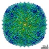 10101MC M: map data used to model this data C: citing same article ( |
|---|---|
| Similar structure data |

-Links
 PDBj
PDBj


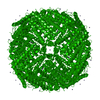
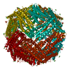
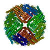
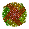
- Assembly
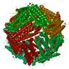
| Deposited unit | 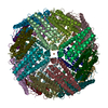
|
|---|---|
| 1 |
|
-Components
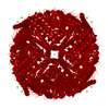
| #1: Protein | Mass: 21097.631 Da / Num. of mol.: 24 Source method: isolated from a genetically manipulated source Source: (gene. exp.)  #2: Chemical | ChemComp-FE /   Mass: 55.845 Da / Num. of mol.: 6 / Source method: obtained synthetically / Formula: Fe Mass: 55.845 Da / Num. of mol.: 6 / Source method: obtained synthetically / Formula: Fe#3: Chemical | ChemComp-ZN /   Mass: 65.409 Da / Num. of mol.: 24 / Source method: isolated from a natural source / Formula: Zn Mass: 65.409 Da / Num. of mol.: 24 / Source method: isolated from a natural source / Formula: Zn#4: Water | ChemComp-HOH / |  Mass: 18.015 Da / Num. of mol.: 3854 / Source method: isolated from a natural source / Formula: H2O Mass: 18.015 Da / Num. of mol.: 3854 / Source method: isolated from a natural source / Formula: H2OHas ligand of interest | N | |
|---|
-Experimental details
-Experiment
| Experiment | Method: ELECTRON MICROSCOPY |
|---|---|
| EM experiment | Aggregation state: PARTICLE / 3D reconstruction method: single particle reconstruction |
- Sample preparation
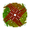
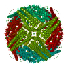
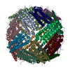
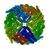
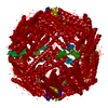
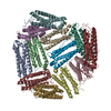
| Component | Name: Octahedral complex of 24 apoferritin chains / Type: COMPLEX / Entity ID: #1 / Source: RECOMBINANT | |||||||||||||||
|---|---|---|---|---|---|---|---|---|---|---|---|---|---|---|---|---|
| Molecular weight | Value: 0.5 MDa / Experimental value: NO | |||||||||||||||
| Source (natural) | Organism: | |||||||||||||||
| Source (recombinant) | Organism: | |||||||||||||||
| Buffer solution | pH: 7.5 | |||||||||||||||
| Buffer component |
| |||||||||||||||
| Specimen | Conc.: 1.5 mg/ml / Embedding applied: NO / Shadowing applied: NO / Staining applied: NO / Vitrification applied: YES | |||||||||||||||
| Vitrification | Instrument: FEI VITROBOT MARK IV / Cryogen name: ETHANE / Humidity: 100 % / Chamber temperature: 283 K |
- Electron microscopy imaging
| Experimental equipment |  Model: Titan Krios / Image courtesy: FEI Company |
|---|---|
| Microscopy | Model: FEI TITAN KRIOS |
| Electron gun | Electron source:  FIELD EMISSION GUN / Accelerating voltage: 300 kV / Illumination mode: FLOOD BEAM FIELD EMISSION GUN / Accelerating voltage: 300 kV / Illumination mode: FLOOD BEAM |
| Electron lens | Mode: BRIGHT FIELD / Nominal magnification: 120000 X / Calibrated magnification: 182927 X / Nominal defocus max: 1500 nm / Nominal defocus min: 500 nm / Calibrated defocus min: 200 nm / Calibrated defocus max: 2000 nm / Cs: 2.7 mm / Alignment procedure: COMA FREE |
| Specimen holder | Cryogen: NITROGEN / Specimen holder model: FEI TITAN KRIOS AUTOGRID HOLDER |
| Image recording | Average exposure time: 30 sec. / Electron dose: 30 e/Å2 / Detector mode: COUNTING / Film or detector model: FEI FALCON III (4k x 4k) / Num. of grids imaged: 1 / Num. of real images: 1788 Details: Some images were discarding after motion correction (MotionCor2) and ctf estimation (CTFfind4), leaving 1745 for processing. |
- Processing
| Software | Name: PHENIX / Version: 1.12_2829: / Classification: refinement | ||||||||||||||||||||||||||||||||
|---|---|---|---|---|---|---|---|---|---|---|---|---|---|---|---|---|---|---|---|---|---|---|---|---|---|---|---|---|---|---|---|---|---|
| EM software |
| ||||||||||||||||||||||||||||||||
| CTF correction | Type: PHASE FLIPPING AND AMPLITUDE CORRECTION | ||||||||||||||||||||||||||||||||
| Particle selection | Num. of particles selected: 477115 | ||||||||||||||||||||||||||||||||
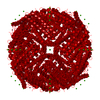
| Symmetry | Point symmetry: O (octahedral) | ||||||||||||||||||||||||||||||||
| 3D reconstruction | Resolution: 1.84 Å / Resolution method: FSC 0.143 CUT-OFF / Num. of particles: 441902 / Symmetry type: POINT | ||||||||||||||||||||||||||||||||
| Atomic model building | Protocol: OTHER / Space: REAL / Details: phenix.real-space-refine | ||||||||||||||||||||||||||||||||
| Atomic model building | PDB-ID: 2CIH Accession code: 2CIH / Source name: PDB / Type: experimental model |

