- EMDB-10714: Apoferritin from horse spleen, prepared using standard Vitrobot w... -
+
Open data
ID or keywords:
Loading...
-
Basic information
Entry
Database: EMDB / ID: EMD-10714
Title
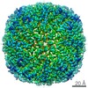
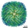
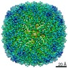
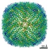
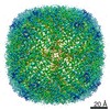
Apoferritin from horse spleen, prepared using standard Vitrobot workflow
Map data
Apoferritin from horse spleen, preparation by Vitrobot
Sample
Complex: Apoferritin from horse spleen
Function / homology
Function and homology information
ferritin complex / autolysosome / ferric iron binding / autophagosome / iron ion transport / ferrous iron binding / cytoplasmic vesicle / intracellular iron ion homeostasis / iron ion binding / cytoplasm Similarity search - Function
Korea, Republic Of, Switzerland, United Kingdom, 7 items
Organization
Grant number
Country
National Research Foundation (NRF, Korea)
NRF-2015K1A1A2033054
Korea, Republic Of
European Research Council (ERC)
Switzerland
Swiss National Science Foundation
Switzerland
Cancer Research UK
United Kingdom
The Francis Crick Institute
United Kingdom
Wellcome Trust
United Kingdom
Medical Research Council (MRC, United Kingdom)
United Kingdom
Citation
Journal: Nat Commun / Year: 2020 Title: Modular microfluidics enables kinetic insight from time-resolved cryo-EM. Authors: Märt-Erik Mäeots / Byungjin Lee / Andrea Nans / Seung-Geun Jeong / Mohammad M N Esfahani / Shan Ding / Daniel J Smith / Chang-Soo Lee / Sung Sik Lee / Matthias Peter / Radoslav I Enchev / Abstract: Mechanistic understanding of biochemical reactions requires structural and kinetic characterization of the underlying chemical processes. However, no single experimental technique can provide this ...Mechanistic understanding of biochemical reactions requires structural and kinetic characterization of the underlying chemical processes. However, no single experimental technique can provide this information in a broadly applicable manner and thus structural studies of static macromolecules are often complemented by biophysical analysis. Moreover, the common strategy of utilizing mutants or crosslinking probes to stabilize intermediates is prone to trapping off-pathway artefacts and precludes determining the order of molecular events. Here we report a time-resolved sample preparation method for cryo-electron microscopy (trEM) using a modular microfluidic device, featuring a 3D-mixing unit and variable delay lines that enables automated, fast, and blot-free sample vitrification. This approach not only preserves high-resolution structural detail but also substantially improves sample integrity and protein distribution across the vitreous ice. We validate the method by visualising reaction intermediates of early RecA filament growth across three orders of magnitude on sub-second timescales. The trEM method reported here is versatile, reproducible, and readily adaptable to a broad spectrum of fundamental questions in biology.
History
Deposition
Feb 28, 2020
-
Header (metadata) release
Jul 22, 2020
-
Map release
Jul 22, 2020
-
Update
Jul 22, 2020
-
Current status
Jul 22, 2020
Processing site: PDBe / Status: Released
-
Structure visualization
Movie
Surface view with section colored by density value
In the structure databanks used in Yorodumi, some data are registered as the other names, "COVID-19 virus" and "2019-nCoV". Here are the details of the virus and the list of structure data.
Jan 31, 2019. EMDB accession codes are about to change! (news from PDBe EMDB page)
EMDB accession codes are about to change! (news from PDBe EMDB page)
The allocation of 4 digits for EMDB accession codes will soon come to an end. Whilst these codes will remain in use, new EMDB accession codes will include an additional digit and will expand incrementally as the available range of codes is exhausted. The current 4-digit format prefixed with “EMD-” (i.e. EMD-XXXX) will advance to a 5-digit format (i.e. EMD-XXXXX), and so on. It is currently estimated that the 4-digit codes will be depleted around Spring 2019, at which point the 5-digit format will come into force.
The EM Navigator/Yorodumi systems omit the EMD- prefix.
Related info.:Q: What is EMD? / ID/Accession-code notation in Yorodumi/EM Navigator
Yorodumi is a browser for structure data from EMDB, PDB, SASBDB, etc.
This page is also the successor to EM Navigator detail page, and also detail information page/front-end page for Omokage search.
The word "yorodu" (or yorozu) is an old Japanese word meaning "ten thousand". "mi" (miru) is to see.
Related info.:EMDB / PDB / SASBDB / Comparison of 3 databanks / Yorodumi Search / Aug 31, 2016. New EM Navigator & Yorodumi / Yorodumi Papers / Jmol/JSmol / Function and homology information / Changes in new EM Navigator and Yorodumi
 Movie
Movie Controller

 Yorodumi
 Open data
 Basic information
 Map data
 Sample
 Function and homology information
 Authors
 Korea, Republic Of,
  Switzerland,
  United Kingdom, 7 items
  Citation
 Structure visualization
 Downloads & links
 emd_10714.png
 http://ftp.pdbj.org/pub/emdb/structures/EMD-10714
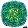


 X (Sec.)
 Y (Row.)
 Z (Col.)


 Sample components
 Processing
 Electron microscopy
 FIELD EMISSION GUN
