 Movie
Movie Controller


+ Open data

- Basic information
| Entry | Database: PDB / ID: 6m4o | |||||||||||||||||||||||||||
|---|---|---|---|---|---|---|---|---|---|---|---|---|---|---|---|---|---|---|---|---|---|---|---|---|---|---|---|---|
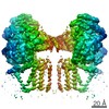
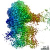
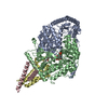
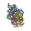
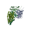
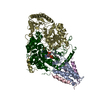
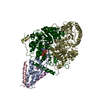
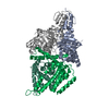
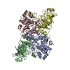
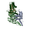
| Title | Cryo-EM structure of the monomeric SPT-ORMDL3 complex | |||||||||||||||||||||||||||
 Components Components |
| |||||||||||||||||||||||||||
 Keywords Keywords | TRANSFERASE / membrane protein / lipid synthesis | |||||||||||||||||||||||||||
| Function / homology |  Function and homology information Function and homology informationsphinganine biosynthetic process / regulation of fat cell apoptotic process / negative regulation of ceramide biosynthetic process / intracellular sphingolipid homeostasis / serine palmitoyltransferase complex / sphingomyelin biosynthetic process / serine C-palmitoyltransferase / serine C-palmitoyltransferase activity / ceramide metabolic process / sphingosine biosynthetic process ...sphinganine biosynthetic process / regulation of fat cell apoptotic process / negative regulation of ceramide biosynthetic process / intracellular sphingolipid homeostasis / serine palmitoyltransferase complex / sphingomyelin biosynthetic process / serine C-palmitoyltransferase / serine C-palmitoyltransferase activity / ceramide metabolic process / sphingosine biosynthetic process / sphingolipid metabolic process / Sphingolipid de novo biosynthesis / regulation of smooth muscle contraction / sphingolipid biosynthetic process / ceramide biosynthetic process / positive regulation of lipophagy / negative regulation of B cell apoptotic process / motor behavior / adipose tissue development / specific granule membrane / myelination / positive regulation of autophagy / secretory granule membrane / positive regulation of protein localization to nucleus / intracellular protein localization / pyridoxal phosphate binding / Neutrophil degranulation / endoplasmic reticulum membrane / endoplasmic reticulum / plasma membrane Similarity search - Function | |||||||||||||||||||||||||||
| Biological species |  Homo sapiens (human) Homo sapiens (human) | |||||||||||||||||||||||||||
| Method | ELECTRON MICROSCOPY / single particle reconstruction / cryo EM / Resolution: 3.4 Å | |||||||||||||||||||||||||||
 Authors Authors | Li, S.S. / Xie, T. / Wang, L. / Gong, X. | |||||||||||||||||||||||||||
 Citation Citation | Journal: Nat Struct Mol Biol / Year: 2021 Title: Structural insights into the assembly and substrate selectivity of human SPT-ORMDL3 complex. Authors: Sisi Li / Tian Xie / Peng Liu / Lei Wang / Xin Gong /  Abstract: Human serine palmitoyltransferase (SPT) complex catalyzes the initial and rate-limiting step in the de novo biosynthesis of all sphingolipids. ORMDLs regulate SPT function, with human ORMDL3 being ...Human serine palmitoyltransferase (SPT) complex catalyzes the initial and rate-limiting step in the de novo biosynthesis of all sphingolipids. ORMDLs regulate SPT function, with human ORMDL3 being related to asthma. Here we report three high-resolution cryo-EM structures: the human SPT complex, composed of SPTLC1, SPTLC2 and SPTssa; the SPT-ORMDL3 complex; and the SPT-ORMDL3 complex bound to two substrates, PLP-L-serine (PLS) and a non-reactive palmitoyl-CoA analogue. SPTLC1 and SPTLC2 form a dimer of heterodimers as the catalytic core. SPTssa participates in acyl-CoA coordination, thereby stimulating the SPT activity and regulating the substrate selectivity. ORMDL3 is located in the center of the complex, serving to stabilize the SPT assembly. Our structural and biochemical analyses provide a molecular basis for the assembly and substrate selectivity of the SPT and SPT-ORMDL3 complexes, and lay a foundation for mechanistic understanding of sphingolipid homeostasis and for related therapeutic drug development. | |||||||||||||||||||||||||||
| History |
|
- Structure visualization
| Movie |
Movie viewer |
|---|---|
| Structure viewer | Molecule: MolmilJmol/JSmol |
- Downloads & links
-Download
| PDBx/mmCIF format | 6m4o.cif.gz | 218.1 KB | Display | PDBx/mmCIF format |
|---|---|---|---|---|
| PDB format | pdb6m4o.ent.gz | 167.3 KB | Display | PDB format |
| PDBx/mmJSON format | 6m4o.json.gz | Tree view | PDBx/mmJSON format | |
| Others |  Other downloads Other downloads |
-Validation report
| Arichive directory | https://data.pdbj.org/pub/pdb/validation_reports/m4/6m4oftp://data.pdbj.org/pub/pdb/validation_reports/m4/6m4o | HTTPS FTP |
|---|
-Related structure data
| Related structure data |  30080MC  6m4nC  7cqiC  7cqkC C: citing same article ( M: map data used to model this data |
|---|---|
| Similar structure data |


-Links
 PDBj
PDBj- Assembly
| Deposited unit | 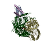
|
|---|---|
| 1 |
|
-Components
| #1: Protein | Mass: 52806.770 Da / Num. of mol.: 2 Source method: isolated from a genetically manipulated source Source: (gene. exp.) Homo sapiens (human) / Gene: SPTLC1, LCB1 / Production host: Homo sapiens (human) / References: UniProt: O15269, serine C-palmitoyltransferase#2: Protein | | Mass: 63004.160 Da / Num. of mol.: 1 Source method: isolated from a genetically manipulated source Source: (gene. exp.) Homo sapiens (human) / Gene: SPTLC2, KIAA0526, LCB2 / Production host: Homo sapiens (human) / References: UniProt: O15270, serine C-palmitoyltransferase#3: Protein | | Mass: 17512.594 Da / Num. of mol.: 1 Source method: isolated from a genetically manipulated source Source: (gene. exp.) Homo sapiens (human) / Gene: ORMDL3 / Production host: Homo sapiens (human) / References: UniProt: Q8N138#4: Protein | | Mass: 10742.409 Da / Num. of mol.: 1 Source method: isolated from a genetically manipulated source Source: (gene. exp.) Homo sapiens (human) / Gene: SPTSSA, C14orf147, SSSPTA / Production host: Homo sapiens (human) / References: UniProt: Q969W0#5: Chemical | ChemComp-PLP / |   Mass: 247.142 Da / Num. of mol.: 1 / Source method: obtained synthetically / Formula: C8H10NO6P / Feature type: SUBJECT OF INVESTIGATION Mass: 247.142 Da / Num. of mol.: 1 / Source method: obtained synthetically / Formula: C8H10NO6P / Feature type: SUBJECT OF INVESTIGATIONHas ligand of interest | Y | Has protein modification | N | |
|---|
-Experimental details
-Experiment
| Experiment | Method: ELECTRON MICROSCOPY |
|---|---|
| EM experiment | Aggregation state: PARTICLE / 3D reconstruction method: single particle reconstruction |
- Sample preparation
| Component | Name: membrane protein 2 / Type: COMPLEX / Entity ID: #1-#4 / Source: RECOMBINANT |
|---|---|
| Source (natural) | Organism: Homo sapiens (human) |
| Source (recombinant) | Organism: Homo sapiens (human) |
| Buffer solution | pH: 8 |
| Specimen | Embedding applied: NO / Shadowing applied: NO / Staining applied: NO / Vitrification applied: YES |
| Vitrification | Cryogen name: ETHANE |
- Electron microscopy imaging
| Experimental equipment |  Model: Titan Krios / Image courtesy: FEI Company |
|---|---|
| Microscopy | Model: FEI TITAN KRIOS |
| Electron gun | Electron source:  FIELD EMISSION GUN / Accelerating voltage: 300 kV / Illumination mode: FLOOD BEAM FIELD EMISSION GUN / Accelerating voltage: 300 kV / Illumination mode: FLOOD BEAM |
| Electron lens | Mode: BRIGHT FIELD |
| Image recording | Electron dose: 50 e/Å2 / Film or detector model: GATAN K2 SUMMIT (4k x 4k) |
- Processing
| CTF correction | Type: NONE |
|---|---|
| 3D reconstruction | Resolution: 3.4 Å / Resolution method: FSC 0.143 CUT-OFF / Num. of particles: 59924 / Symmetry type: POINT |
