 ムービー
ムービー コントローラー


+ データを開く

- 基本情報
基本情報
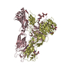
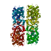
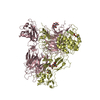
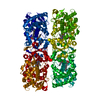
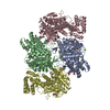
| 登録情報 | データベース: PDB / ID: 1poi | ||||||
|---|---|---|---|---|---|---|---|
| タイトル | CRYSTAL STRUCTURE OF GLUTACONATE COENZYME A-TRANSFERASE FROM ACIDAMINOCOCCUS FERMENTANS TO 2.55 ANGSTOMS RESOLUTION | ||||||
 要素 要素 | (GLUTACONATE COENZYME A-TRANSFERASE) x 2 | ||||||
 キーワード キーワード | TRANSFERASE / COA / GLUTAMATE / PROTEIN FERMENTATION | ||||||
| 機能・相同性 |  機能・相同性情報 機能・相同性情報 | ||||||
| 生物種 |  Acidaminococcus fermentans (バクテリア) Acidaminococcus fermentans (バクテリア) | ||||||
| 手法 |  X線回折 / 多重同系置換 / 解像度: 2.5 Å X線回折 / 多重同系置換 / 解像度: 2.5 Å | ||||||
 データ登録者 データ登録者 | Jacob, U. / Mack, M. / Clausen, T. / Huber, R. / Buckel, W. / Messerschmidt, A. | ||||||
 引用 引用 | ジャーナル: Structure / 年: 1997 タイトル: Glutaconate CoA-transferase from Acidaminococcus fermentans: the crystal structure reveals homology with other CoA-transferases. 著者: Jacob, U. / Mack, M. / Clausen, T. / Huber, R. / Buckel, W. / Messerschmidt, A. | ||||||
| 履歴 |
|
- 構造の表示
構造の表示
| 構造ビューア | 分子: MolmilJmol/JSmol |
|---|
- ダウンロードとリンク
-ダウンロード
| PDBx/mmCIF形式 | 1poi.cif.gz | 238 KB | 表示 | PDBx/mmCIF形式 |
|---|---|---|---|---|
| PDB形式 | pdb1poi.ent.gz | 191.1 KB | 表示 | PDB形式 |
| PDBx/mmJSON形式 | 1poi.json.gz | ツリー表示 | PDBx/mmJSON形式 | |
| その他 |  その他のダウンロード その他のダウンロード |
-検証レポート
| アーカイブディレクトリ | https://data.pdbj.org/pub/pdb/validation_reports/po/1poiftp://data.pdbj.org/pub/pdb/validation_reports/po/1poi | HTTPS FTP |
|---|
-関連構造データ


-リンク
 PDBj
PDBj- 集合体
集合体
| 登録構造単位 | 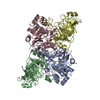
| ||||||||||||
|---|---|---|---|---|---|---|---|---|---|---|---|---|---|
| 1 | 
| ||||||||||||
| 単位格子 |
| ||||||||||||
| Components on special symmetry positions |
| ||||||||||||
| 非結晶学的対称性 (NCS) | NCS oper:
|
-要素
| #1: タンパク質 | 分子量: 35390.273 Da / 分子数: 2 / 由来タイプ: 組換発現 由来: (組換発現) Acidaminococcus fermentans (バクテリア)細胞内の位置 (発現宿主): CYTOSOL / 発現宿主: #2: タンパク質 | 分子量: 28566.547 Da / 分子数: 2 / 由来タイプ: 組換発現 由来: (組換発現) Acidaminococcus fermentans (バクテリア)細胞内の位置 (発現宿主): CYTOSOL / 発現宿主: #3: 化合物 | ChemComp-CU / |   分子量: 63.546 Da / 分子数: 1 / 由来タイプ: 合成 / 式: Cu 分子量: 63.546 Da / 分子数: 1 / 由来タイプ: 合成 / 式: Cu#4: 水 | ChemComp-HOH / |  分子量: 18.015 Da / 分子数: 438 / 由来タイプ: 天然 / 式: H2O 分子量: 18.015 Da / 分子数: 438 / 由来タイプ: 天然 / 式: H2O |
|---|
-実験情報
-実験
| 実験 | 手法: X線回折 / 使用した結晶の数: 2 |
|---|
- 試料調製
試料調製
| 結晶 | マシュー密度: 2.8 Å3/Da / 溶媒含有率: 56 % | ||||||||||||||||||||||||||||||||||||||||||||||||||||||||||||
|---|---|---|---|---|---|---|---|---|---|---|---|---|---|---|---|---|---|---|---|---|---|---|---|---|---|---|---|---|---|---|---|---|---|---|---|---|---|---|---|---|---|---|---|---|---|---|---|---|---|---|---|---|---|---|---|---|---|---|---|---|---|
| 結晶化 | pH: 5.5 / 詳細: AMMONIUM SULFATE/PEG PH=5.5 | ||||||||||||||||||||||||||||||||||||||||||||||||||||||||||||
| 結晶化 | *PLUS 温度: 20 ℃ / 手法: 蒸気拡散法, シッティングドロップ法 | ||||||||||||||||||||||||||||||||||||||||||||||||||||||||||||
| 溶液の組成 | *PLUS
|
-データ収集
| 回折 | 平均測定温度: 298 K |
|---|---|
| 放射光源 | 波長: 1.5418 |
| 検出器 | タイプ: MARRESEARCH / 検出器: IMAGE PLATE / 日付: 1995年5月1日 |
| 放射 | 単色(M)・ラウエ(L): M / 散乱光タイプ: x-ray |
| 放射波長 | 波長: 1.5418 Å / 相対比: 1 |
| 反射 | 解像度: 2.5→7 Å / Num. obs: 37201 / % possible obs: 83 % / Observed criterion σ(I): 0 / 冗長度: 2.8 % / Rmerge(I) obs: 0.098 |
| 反射 シェル | 解像度: 2.55→2.64 Å / % possible all: 32 |
- 解析
解析
| ソフトウェア |
| ||||||||||||||||||||||||||||||||||||||||||||||||||||||||||||
|---|---|---|---|---|---|---|---|---|---|---|---|---|---|---|---|---|---|---|---|---|---|---|---|---|---|---|---|---|---|---|---|---|---|---|---|---|---|---|---|---|---|---|---|---|---|---|---|---|---|---|---|---|---|---|---|---|---|---|---|---|---|
| 精密化 | 構造決定の手法: 多重同系置換 / 解像度: 2.5→7 Å / Rfactor Rwork: 0.19 / Rfactor obs: 0.19 / 交差検証法: FREE-R / σ(F): 0 | ||||||||||||||||||||||||||||||||||||||||||||||||||||||||||||
| 原子変位パラメータ | Biso mean: 24 Å2 | ||||||||||||||||||||||||||||||||||||||||||||||||||||||||||||
| Refine analyze | Luzzati coordinate error obs: 0.31 Å | ||||||||||||||||||||||||||||||||||||||||||||||||||||||||||||
| 精密化ステップ | サイクル: LAST / 解像度: 2.5→7 Å
| ||||||||||||||||||||||||||||||||||||||||||||||||||||||||||||
| 拘束条件 |
| ||||||||||||||||||||||||||||||||||||||||||||||||||||||||||||
| ソフトウェア | *PLUS 名称: X-PLOR / バージョン: 3.1 / 分類: refinement | ||||||||||||||||||||||||||||||||||||||||||||||||||||||||||||
| 精密化 | *PLUS Num. reflection obs: 37201 / Rfactor Rfree: 0.27 | ||||||||||||||||||||||||||||||||||||||||||||||||||||||||||||
| 溶媒の処理 | *PLUS | ||||||||||||||||||||||||||||||||||||||||||||||||||||||||||||
| 原子変位パラメータ | *PLUS |
