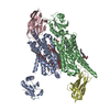
 Movie
Movie Controller


+ Open data

- Basic information
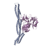
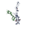
| Entry | Database: PDB / ID: 6m3r | ||||||
|---|---|---|---|---|---|---|---|
| Title | Crystal structure of AnkG/beta4-spectrin complex | ||||||
 Components Components |
| ||||||
 Keywords Keywords | STRUCTURAL PROTEIN / PROTEIN BINDING | ||||||
| Function / homology |  Function and homology information Function and homology informationInteraction between L1 and Ankyrins / regulation of protein targeting / positive regulation of cell communication by electrical coupling / positive regulation of membrane depolarization during cardiac muscle cell action potential / maintenance of protein location in plasma membrane / positive regulation of sodium ion import across plasma membrane / NCAM signaling for neurite out-growth / membrane assembly / protein localization to axon / clustering of voltage-gated sodium channels ...Interaction between L1 and Ankyrins / regulation of protein targeting / positive regulation of cell communication by electrical coupling / positive regulation of membrane depolarization during cardiac muscle cell action potential / maintenance of protein location in plasma membrane / positive regulation of sodium ion import across plasma membrane / NCAM signaling for neurite out-growth / membrane assembly / protein localization to axon / clustering of voltage-gated sodium channels / spectrin / negative regulation of delayed rectifier potassium channel activity / spectrin-associated cytoskeleton / establishment or maintenance of microtubule cytoskeleton polarity / central nervous system projection neuron axonogenesis / regulation of potassium ion transport / COPI-mediated anterograde transport / magnesium ion homeostasis / plasma membrane organization / RAF/MAP kinase cascade / positive regulation of membrane potential / channel activator activity / maintenance of protein location in cell / phosphorylation-dependent protein binding / cell body fiber / cardiac conduction / positive regulation of homotypic cell-cell adhesion / paranode region of axon / axon hillock / positive regulation of sodium ion transport / costamere / negative regulation of endocytosis / cellular response to magnesium ion / axon initial segment / anterograde axonal transport / Golgi to plasma membrane protein transport / juxtaparanode region of axon / regulation of modification of postsynaptic structure / positive regulation of multicellular organism growth / actin filament capping / node of Ranvier / fertilization / adult walking behavior / negative regulation of heart rate / ankyrin binding / cortical actin cytoskeleton / spectrin binding / neuromuscular process / axon development / response to immobilization stress / neuromuscular junction development / adult behavior / mitotic cytokinesis / transmission of nerve impulse / intercalated disc / lateral plasma membrane / sodium channel regulator activity / bicellular tight junction / positive regulation of protein targeting to membrane / phosphatase binding / regulation of sodium ion transport / neuronal action potential / cytoskeletal protein binding / cell projection / axon cytoplasm / axonogenesis / T-tubule / basal plasma membrane / axon guidance / sarcoplasmic reticulum / protein localization to plasma membrane / neuromuscular junction / establishment of protein localization / sensory perception of sound / positive regulation of non-canonical NF-kappaB signal transduction / PML body / phospholipid binding / sarcolemma / synapse organization / structural constituent of cytoskeleton / Z disc / nuclear matrix / actin filament binding / cell junction / intracellular protein localization / actin binding / actin cytoskeleton organization / protein-containing complex assembly / basolateral plasma membrane / cytoskeleton / RNA polymerase II-specific DNA-binding transcription factor binding / transmembrane transporter binding / protein-macromolecule adaptor activity / postsynaptic membrane / lysosome / neuron projection / postsynaptic density / postsynapse / cadherin binding / axon Similarity search - Function | ||||||
| Biological species |  | ||||||
| Method |  X-RAY DIFFRACTION / SYNCHROTRON / MOLECULAR REPLACEMENT / Resolution: 4.313 Å X-RAY DIFFRACTION / SYNCHROTRON / MOLECULAR REPLACEMENT / Resolution: 4.313 Å | ||||||
 Authors Authors | Li, J. / Chen, K. / Zhu, R. / Zhang, M. | ||||||
| Funding support |  Hong Kong, 1items Hong Kong, 1items
| ||||||
 Citation Citation | Journal: J.Mol.Biol. / Year: 2020 Title: Structural Basis Underlying Strong Interactions between Ankyrins and Spectrins. Authors: Li, J. / Chen, K. / Zhu, R. / Zhang, M. | ||||||
| History |
|
- Structure visualization
| Structure viewer | Molecule: MolmilJmol/JSmol |
|---|
- Downloads & links
-Download
| PDBx/mmCIF format | 6m3r.cif.gz | 148.8 KB | Display | PDBx/mmCIF format |
|---|---|---|---|---|
| PDB format | pdb6m3r.ent.gz | 108.5 KB | Display | PDB format |
| PDBx/mmJSON format | 6m3r.json.gz | Tree view | PDBx/mmJSON format | |
| Others |  Other downloads Other downloads |
-Validation report
| Arichive directory | https://data.pdbj.org/pub/pdb/validation_reports/m3/6m3rftp://data.pdbj.org/pub/pdb/validation_reports/m3/6m3r | HTTPS FTP |
|---|
-Related structure data
| Related structure data |  6m3pC 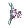 6m3qC 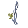 3kbtS  4d8oS S: Starting model for refinement C: citing same article ( |
|---|---|
| Similar structure data |


-Links
 PDBj
PDBj


- Assembly
| Deposited unit | 
| ||||||||
|---|---|---|---|---|---|---|---|---|---|
| 1 |
| ||||||||
| Unit cell |
|
-Components
| #1: Protein | Mass: 54560.531 Da / Num. of mol.: 1 Source method: isolated from a genetically manipulated source Source: (gene. exp.)  |
|---|---|
| #2: Protein | Mass: 36877.199 Da / Num. of mol.: 1 Source method: isolated from a genetically manipulated source Source: (gene. exp.) |
-Experimental details
-Experiment
| Experiment | Method: X-RAY DIFFRACTION / Number of used crystals: 1 |
|---|
- Sample preparation
| Crystal | Density Matthews: 6.5 Å3/Da / Density % sol: 81.08 % |
|---|---|
| Crystal grow | Temperature: 289 K / Method: vapor diffusion / pH: 8.5 Details: 0.8 M potassium sodium tartrate tetrahydrate, 0.1 M Tris, pH 8.5 and 0.5% w/v polyethylene glycol monomethyl ether 5,000 |
-Data collection
| Diffraction | Mean temperature: 100 K / Serial crystal experiment: N | |||||||||||||||||||||||||||||||||||||||||||||||||||||||||||||||||||||||||||||||||||||||||||||||||||||||||||||||||||||||||||||||||||||||||||||||||||||||||||||||||||||||||||||||||||||||||||||
|---|---|---|---|---|---|---|---|---|---|---|---|---|---|---|---|---|---|---|---|---|---|---|---|---|---|---|---|---|---|---|---|---|---|---|---|---|---|---|---|---|---|---|---|---|---|---|---|---|---|---|---|---|---|---|---|---|---|---|---|---|---|---|---|---|---|---|---|---|---|---|---|---|---|---|---|---|---|---|---|---|---|---|---|---|---|---|---|---|---|---|---|---|---|---|---|---|---|---|---|---|---|---|---|---|---|---|---|---|---|---|---|---|---|---|---|---|---|---|---|---|---|---|---|---|---|---|---|---|---|---|---|---|---|---|---|---|---|---|---|---|---|---|---|---|---|---|---|---|---|---|---|---|---|---|---|---|---|---|---|---|---|---|---|---|---|---|---|---|---|---|---|---|---|---|---|---|---|---|---|---|---|---|---|---|---|---|---|---|---|---|
| Diffraction source | Source: SYNCHROTRON / Site: SSRF / Beamline: BL19U1 / Wavelength: 0.97853 Å | |||||||||||||||||||||||||||||||||||||||||||||||||||||||||||||||||||||||||||||||||||||||||||||||||||||||||||||||||||||||||||||||||||||||||||||||||||||||||||||||||||||||||||||||||||||||||||||
| Detector | Type: DECTRIS PILATUS3 6M / Detector: PIXEL / Date: Jun 20, 2016 | |||||||||||||||||||||||||||||||||||||||||||||||||||||||||||||||||||||||||||||||||||||||||||||||||||||||||||||||||||||||||||||||||||||||||||||||||||||||||||||||||||||||||||||||||||||||||||||
| Radiation | Protocol: SINGLE WAVELENGTH / Monochromatic (M) / Laue (L): M / Scattering type: x-ray | |||||||||||||||||||||||||||||||||||||||||||||||||||||||||||||||||||||||||||||||||||||||||||||||||||||||||||||||||||||||||||||||||||||||||||||||||||||||||||||||||||||||||||||||||||||||||||||
| Radiation wavelength | Wavelength: 0.97853 Å / Relative weight: 1 | |||||||||||||||||||||||||||||||||||||||||||||||||||||||||||||||||||||||||||||||||||||||||||||||||||||||||||||||||||||||||||||||||||||||||||||||||||||||||||||||||||||||||||||||||||||||||||||
| Reflection | Resolution: 4.3→50 Å / Num. obs: 16538 / % possible obs: 99.9 % / Redundancy: 15.7 % / Biso Wilson estimate: 77.93 Å2 / Rmerge(I) obs: 0.159 / Rpim(I) all: 0.059 / Rrim(I) all: 0.142 / Χ2: 0.961 / Net I/σ(I): 2.2 / Num. measured all: 259971 | |||||||||||||||||||||||||||||||||||||||||||||||||||||||||||||||||||||||||||||||||||||||||||||||||||||||||||||||||||||||||||||||||||||||||||||||||||||||||||||||||||||||||||||||||||||||||||||
| Reflection shell | Diffraction-ID: 1
|
- Processing
| Software |
| ||||||||||||||||||||||||||||||||||||
|---|---|---|---|---|---|---|---|---|---|---|---|---|---|---|---|---|---|---|---|---|---|---|---|---|---|---|---|---|---|---|---|---|---|---|---|---|---|
| Refinement | Method to determine structure: MOLECULAR REPLACEMENT Starting model: 4D8O, 3KBT Resolution: 4.313→39.896 Å / SU ML: 0.74 / Cross valid method: THROUGHOUT / σ(F): 1.34 / Phase error: 31.9
| ||||||||||||||||||||||||||||||||||||
| Solvent computation | Shrinkage radii: 0.9 Å / VDW probe radii: 1.11 Å | ||||||||||||||||||||||||||||||||||||
| Displacement parameters | Biso max: 349.62 Å2 / Biso mean: 87.6322 Å2 / Biso min: 3.08 Å2 | ||||||||||||||||||||||||||||||||||||
| Refinement step | Cycle: final / Resolution: 4.313→39.896 Å
| ||||||||||||||||||||||||||||||||||||
| Refine LS restraints |
| ||||||||||||||||||||||||||||||||||||
| LS refinement shell | Refine-ID: X-RAY DIFFRACTION / Rfactor Rfree error: 0
|
