 Movie
Movie Controller


+ Open data

- Basic information
| Entry | Database: PDB / ID: 6m3p | ||||||
|---|---|---|---|---|---|---|---|
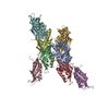
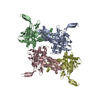
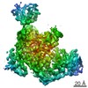
| Title | Crystal structure of AnkG/beta2-spectrin complex | ||||||
 Components Components |
| ||||||
 Keywords Keywords | STRUCTURAL PROTEIN / PROTEIN BINDING | ||||||
| Function / homology |  Function and homology information Function and homology informationInteraction between L1 and Ankyrins / regulation of protein targeting / positive regulation of cell communication by electrical coupling / positive regulation of membrane depolarization during cardiac muscle cell action potential / maintenance of protein location in plasma membrane / positive regulation of sodium ion import across plasma membrane / regulation of SMAD protein signal transduction / RHOV GTPase cycle / NCAM signaling for neurite out-growth / membrane assembly ...Interaction between L1 and Ankyrins / regulation of protein targeting / positive regulation of cell communication by electrical coupling / positive regulation of membrane depolarization during cardiac muscle cell action potential / maintenance of protein location in plasma membrane / positive regulation of sodium ion import across plasma membrane / regulation of SMAD protein signal transduction / RHOV GTPase cycle / NCAM signaling for neurite out-growth / membrane assembly / central nervous system formation / RHOU GTPase cycle / protein localization to axon / clustering of voltage-gated sodium channels / spectrin / negative regulation of delayed rectifier potassium channel activity / spectrin-associated cytoskeleton / establishment or maintenance of microtubule cytoskeleton polarity / regulation of potassium ion transport / cuticular plate / COPI-mediated anterograde transport / magnesium ion homeostasis / plasma membrane organization / RAF/MAP kinase cascade / positive regulation of membrane potential / channel activator activity / maintenance of protein location in cell / phosphorylation-dependent protein binding / positive regulation of homotypic cell-cell adhesion / paranode region of axon / positive regulation of sodium ion transport / costamere / negative regulation of endocytosis / cellular response to magnesium ion / axon initial segment / anterograde axonal transport / Golgi to plasma membrane protein transport / regulation of modification of postsynaptic structure / actin filament capping / node of Ranvier / M band / ankyrin binding / cortical actin cytoskeleton / spectrin binding / axon development / response to immobilization stress / neuromuscular junction development / cortical cytoskeleton / mitotic cytokinesis / intercalated disc / lateral plasma membrane / sodium channel regulator activity / bicellular tight junction / positive regulation of protein targeting to membrane / axolemma / positive regulation of interleukin-2 production / regulation of protein localization to plasma membrane / neuronal action potential / cytoskeletal protein binding / cell projection / axon cytoplasm / endomembrane system / axonogenesis / T-tubule / basal plasma membrane / axon guidance / sarcoplasmic reticulum / central nervous system development / protein localization to plasma membrane / positive regulation of protein localization to plasma membrane / neuromuscular junction / establishment of protein localization / positive regulation of non-canonical NF-kappaB signal transduction / sarcolemma / phospholipid binding / synapse organization / structural constituent of cytoskeleton / Z disc / actin filament binding / cell junction / actin cytoskeleton organization / GTPase binding / basolateral plasma membrane / cytoskeleton / RNA polymerase II-specific DNA-binding transcription factor binding / transmembrane transporter binding / protein-macromolecule adaptor activity / calmodulin binding / postsynaptic membrane / lysosome / neuron projection / postsynaptic density / postsynapse / cadherin binding / axon / positive regulation of gene expression / regulation of transcription by RNA polymerase II / synapse / dendrite / protein-containing complex binding Similarity search - Function | ||||||
| Biological species |  | ||||||
| Method |  X-RAY DIFFRACTION / SYNCHROTRON / MOLECULAR REPLACEMENT / Resolution: 3.312 Å X-RAY DIFFRACTION / SYNCHROTRON / MOLECULAR REPLACEMENT / Resolution: 3.312 Å | ||||||
 Authors Authors | Li, J. / Chen, K. / Zhu, R. / Zhang, M. | ||||||
| Funding support |  Hong Kong, 1items Hong Kong, 1items
| ||||||
 Citation Citation | Journal: J.Mol.Biol. / Year: 2020 Title: Structural Basis Underlying Strong Interactions between Ankyrins and Spectrins. Authors: Li, J. / Chen, K. / Zhu, R. / Zhang, M. | ||||||
| History |
|
- Structure visualization
| Structure viewer | Molecule: MolmilJmol/JSmol |
|---|
- Downloads & links
-Download
| PDBx/mmCIF format | 6m3p.cif.gz | 283.9 KB | Display | PDBx/mmCIF format |
|---|---|---|---|---|
| PDB format | pdb6m3p.ent.gz | 213.8 KB | Display | PDB format |
| PDBx/mmJSON format | 6m3p.json.gz | Tree view | PDBx/mmJSON format | |
| Others |  Other downloads Other downloads |
-Validation report
| Arichive directory | https://data.pdbj.org/pub/pdb/validation_reports/m3/6m3pftp://data.pdbj.org/pub/pdb/validation_reports/m3/6m3p | HTTPS FTP |
|---|
-Related structure data
| Related structure data | 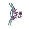 6m3qC 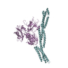 6m3rC 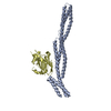 3kbtS  4d8oS S: Starting model for refinement C: citing same article ( |
|---|---|
| Similar structure data |


-Links
 PDBj
PDBj


- Assembly
| Deposited unit | 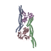
| |||||||||||||||||||||||||||||||||||||||||||||||||||||||||||||||||||||||||||||||||||||||||||||||||||||||||||||
|---|---|---|---|---|---|---|---|---|---|---|---|---|---|---|---|---|---|---|---|---|---|---|---|---|---|---|---|---|---|---|---|---|---|---|---|---|---|---|---|---|---|---|---|---|---|---|---|---|---|---|---|---|---|---|---|---|---|---|---|---|---|---|---|---|---|---|---|---|---|---|---|---|---|---|---|---|---|---|---|---|---|---|---|---|---|---|---|---|---|---|---|---|---|---|---|---|---|---|---|---|---|---|---|---|---|---|---|---|---|---|
| 1 | 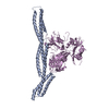
| |||||||||||||||||||||||||||||||||||||||||||||||||||||||||||||||||||||||||||||||||||||||||||||||||||||||||||||
| 2 | 
| |||||||||||||||||||||||||||||||||||||||||||||||||||||||||||||||||||||||||||||||||||||||||||||||||||||||||||||
| Unit cell |
| |||||||||||||||||||||||||||||||||||||||||||||||||||||||||||||||||||||||||||||||||||||||||||||||||||||||||||||
| Noncrystallographic symmetry (NCS) | NCS domain:
NCS domain segments:
|