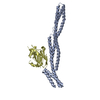
- PDB-3kbt: Crystal structure of the ankyrin binding domain of human erythroi... -
+
Open data
ID or keywords:
Loading...
-
Basic information
Entry
Database: PDB / ID: 3kbt
Title
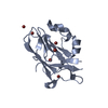
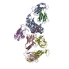
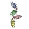
Crystal structure of the ankyrin binding domain of human erythroid beta spectrin (repeats 13-15) in complex with the spectrin binding domain of human erythroid ankyrin (ZU5-ANK)
Mass: 37245.320 Da / Num. of mol.: 2 / Fragment: UNP residues 1583-1906 Source method: isolated from a genetically manipulated source Source: (gene. exp.) Homo sapiens (human) / Gene: SPTB, SPTB1 / Plasmid: pMCSG7 / Production host: Escherichia coli (E. coli) / Strain (production host): BL21(DE3) / References: UniProt: P11277
#2: Protein
Ankyrin-1 / Erythrocyte ankyrin / Ankyrin-R
Mass: 17885.594 Da / Num. of mol.: 2 / Fragment: UNP residues 911-1068 Source method: isolated from a genetically manipulated source Source: (gene. exp.) Homo sapiens (human) / Gene: ANK, ANK1 / Plasmid: pICANTE / Production host: Escherichia coli (E. coli) / Strain (production host): ER2566 / References: UniProt: P16157
Sequence details
EL TO DV SEQUENCE CONFLICT IN UNP ENTRY P16157
-
Experimental details
-
Experiment
Experiment
Method: X-RAY DIFFRACTION / Number of used crystals: 1
-
Sample preparation
Crystal
Density Matthews: 2.71 Å3/Da / Density % sol: 54.54 %
Crystal grow
Temperature: 283 K / Method: vapor diffusion, hanging drop / pH: 6.5 Details: Protein complex at 7.5 mg/mL mixed 1:1 with 0.05 M MES pH 6.5, 0.2 M ammonium acetate, 0.01 M calcium chloride, 10% PEG-4000, 5 mM dithiothreitol, VAPOR DIFFUSION, HANGING DROP, temperature 283K
In the structure databanks used in Yorodumi, some data are registered as the other names, "COVID-19 virus" and "2019-nCoV". Here are the details of the virus and the list of structure data.
Jan 31, 2019. EMDB accession codes are about to change! (news from PDBe EMDB page)
EMDB accession codes are about to change! (news from PDBe EMDB page)
The allocation of 4 digits for EMDB accession codes will soon come to an end. Whilst these codes will remain in use, new EMDB accession codes will include an additional digit and will expand incrementally as the available range of codes is exhausted. The current 4-digit format prefixed with “EMD-” (i.e. EMD-XXXX) will advance to a 5-digit format (i.e. EMD-XXXXX), and so on. It is currently estimated that the 4-digit codes will be depleted around Spring 2019, at which point the 5-digit format will come into force.
The EM Navigator/Yorodumi systems omit the EMD- prefix.
Related info.:Q: What is EMD? / ID/Accession-code notation in Yorodumi/EM Navigator
Yorodumi is a browser for structure data from EMDB, PDB, SASBDB, etc.
This page is also the successor to EM Navigator detail page, and also detail information page/front-end page for Omokage search.
The word "yorodu" (or yorozu) is an old Japanese word meaning "ten thousand". "mi" (miru) is to see.
Related info.:EMDB / PDB / SASBDB / Comparison of 3 databanks / Yorodumi Search / Aug 31, 2016. New EM Navigator & Yorodumi / Yorodumi Papers / Jmol/JSmol / Function and homology information / Changes in new EM Navigator and Yorodumi
 Movie
Movie Controller

 Yorodumi
 Open data
 Basic information
 Components
 Keywords
 Function and homology information
 Homo sapiens (human)
 X-RAY DIFFRACTION /
  Authors
 Citation
 Structure visualization
 Downloads & links
 Other downloads


 PDBj
PDBj


 Assembly
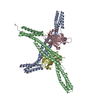


 Sample preparation
 / Beamline: 21-ID-F / Wavelength: 0.97872 Å
 Processing