 Movie
Movie Controller

[English] 日本語
 Yorodumi
- PDB-3kbu: Crystal structure of the ankyrin binding domain of human erythroi... -
+ Open data

- Basic information
| Entry | Database: PDB / ID: 3kbu | ||||||
|---|---|---|---|---|---|---|---|
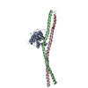
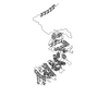
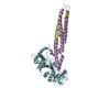
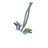
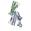
| Title | Crystal structure of the ankyrin binding domain of human erythroid beta spectrin (repeats 13-15) in complex with the spectrin binding domain of human erythroid ankyrin (ZU5-ANK), EMTS derivative | ||||||
 Components Components |
| ||||||
 Keywords Keywords | STRUCTURAL PROTEIN / complex / spectrin / spectrin repeat / three helix bundle / ankyrin binding / disease mutation / ankyrin / ZU5 / beta sandwich / spectrin binding / cytoskeleton / membrane skeleton / Actin capping / Actin-binding / Elliptocytosis / Hereditary hemolytic anemia / Phosphoprotein / Alternative promoter usage / ANK repeat / Lipoprotein / Membrane / Sarcoplasmic reticulum | ||||||
| Function / homology |  Function and homology information Function and homology informationspectrin / spectrin-associated cytoskeleton / maintenance of epithelial cell apical/basal polarity / NrCAM interactions / Neurofascin interactions / ankyrin-1 complex / modification of postsynaptic actin cytoskeleton / CHL1 interactions / cytoskeletal adaptor activity / actin filament capping ...spectrin / spectrin-associated cytoskeleton / maintenance of epithelial cell apical/basal polarity / NrCAM interactions / Neurofascin interactions / ankyrin-1 complex / modification of postsynaptic actin cytoskeleton / CHL1 interactions / cytoskeletal adaptor activity / actin filament capping / M band / Interaction between L1 and Ankyrins / ankyrin binding / cortical actin cytoskeleton / spectrin binding / exocytosis / axolemma / endoplasmic reticulum to Golgi vesicle-mediated transport / COPI-mediated anterograde transport / cell projection / cytoskeleton organization / NCAM signaling for neurite out-growth / protein localization to plasma membrane / sarcoplasmic reticulum / sarcolemma / structural constituent of cytoskeleton / Z disc / cytoplasmic side of plasma membrane / cell junction / actin filament binding / actin cytoskeleton / ATPase binding / RAF/MAP kinase cascade / actin cytoskeleton organization / actin binding / protein phosphatase binding / basolateral plasma membrane / cytoskeleton / transmembrane transporter binding / postsynaptic membrane / postsynapse / neuron projection / glutamatergic synapse / structural molecule activity / enzyme binding / cell surface / signal transduction / protein-containing complex / plasma membrane / cytosol Similarity search - Function | ||||||
| Biological species |  Homo sapiens (human) Homo sapiens (human) | ||||||
| Method |  X-RAY DIFFRACTION / SYNCHROTRON / SAD / Resolution: 2.75 Å X-RAY DIFFRACTION / SYNCHROTRON / SAD / Resolution: 2.75 Å | ||||||
 Authors Authors | Ipsaro, J.J. / Mondragon, A. | ||||||
 Citation Citation | Journal: Blood / Year: 2010 Title: Structural basis for spectrin recognition by ankyrin. Authors: Ipsaro, J.J. / Mondragon, A. | ||||||
| History |
|
- Structure visualization
| Structure viewer | Molecule: MolmilJmol/JSmol |
|---|
- Downloads & links
-Download
| PDBx/mmCIF format | 3kbu.cif.gz | 188 KB | Display | PDBx/mmCIF format |
|---|---|---|---|---|
| PDB format | pdb3kbu.ent.gz | 148.9 KB | Display | PDB format |
| PDBx/mmJSON format | 3kbu.json.gz | Tree view | PDBx/mmJSON format | |
| Others |  Other downloads Other downloads |
-Validation report
| Arichive directory | https://data.pdbj.org/pub/pdb/validation_reports/kb/3kbuftp://data.pdbj.org/pub/pdb/validation_reports/kb/3kbu | HTTPS FTP |
|---|
-Related structure data
| Related structure data | 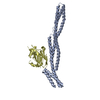 3kbtC  3f57S 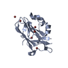 3f59S C: citing same article ( S: Starting model for refinement |
|---|---|
| Similar structure data |


-Links
 PDBj
PDBj


- Assembly
| Deposited unit | 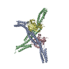
| |||||||||||||||||||||||||||||||||||||||||||||||||||||||||||||||||||||||||||||||||||||||||||||||||||||||||
|---|---|---|---|---|---|---|---|---|---|---|---|---|---|---|---|---|---|---|---|---|---|---|---|---|---|---|---|---|---|---|---|---|---|---|---|---|---|---|---|---|---|---|---|---|---|---|---|---|---|---|---|---|---|---|---|---|---|---|---|---|---|---|---|---|---|---|---|---|---|---|---|---|---|---|---|---|---|---|---|---|---|---|---|---|---|---|---|---|---|---|---|---|---|---|---|---|---|---|---|---|---|---|---|---|---|---|
| 1 | 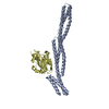
| |||||||||||||||||||||||||||||||||||||||||||||||||||||||||||||||||||||||||||||||||||||||||||||||||||||||||
| 2 | 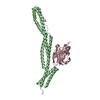
| |||||||||||||||||||||||||||||||||||||||||||||||||||||||||||||||||||||||||||||||||||||||||||||||||||||||||
| Unit cell |
| |||||||||||||||||||||||||||||||||||||||||||||||||||||||||||||||||||||||||||||||||||||||||||||||||||||||||
| Noncrystallographic symmetry (NCS) | NCS domain:
NCS domain segments:
NCS ensembles :
|
-Components
| #1: Protein | Mass: 37219.352 Da / Num. of mol.: 2 / Fragment: UNP residues 1583-1906 / Mutation: E1680C Source method: isolated from a genetically manipulated source Source: (gene. exp.) Homo sapiens (human) / Gene: SPTB, SPTB1 / Plasmid: pMCSG7 / Production host:  #2: Protein | Mass: 17885.594 Da / Num. of mol.: 2 / Fragment: UNP residues 911-1068 Source method: isolated from a genetically manipulated source Source: (gene. exp.) Homo sapiens (human) / Gene: ANK, ANK1 / Plasmid: pICANTE / Production host: #3: Chemical | ChemComp-HG /   Mass: 200.590 Da / Num. of mol.: 8 / Source method: obtained synthetically / Formula: Hg Mass: 200.590 Da / Num. of mol.: 8 / Source method: obtained synthetically / Formula: HgSequence details | EL TO DV SEQUENCE CONFLICT IN UNP ENTRY P16157 | |
|---|
-Experimental details
-Experiment
| Experiment | Method: X-RAY DIFFRACTION / Number of used crystals: 1 |
|---|
- Sample preparation
| Crystal | Density Matthews: 2.78 Å3/Da / Density % sol: 55.74 % |
|---|---|
| Crystal grow | Temperature: 283 K / Method: vapor diffusion, hanging drop / pH: 6.5 Details: Protein complex at 7.5 mg/mL mixed 1:1 with 0.05 M MES pH 6.5, 0.2 M ammonium acetate, 0.01 M calcium chloride, 10% PEG-4000, VAPOR DIFFUSION, HANGING DROP, temperature 283K |
-Data collection
| Diffraction | Mean temperature: 100 K |
|---|---|
| Diffraction source | Source: SYNCHROTRON / Site: APS  / Beamline: 21-ID-F / Wavelength: 0.97872 Å / Beamline: 21-ID-F / Wavelength: 0.97872 Å |
| Detector | Type: RAYONIX MX-225 / Detector: CCD / Date: Nov 7, 2008 |
| Radiation | Monochromator: Diamond laue / Protocol: SINGLE WAVELENGTH / Monochromatic (M) / Laue (L): M / Scattering type: x-ray |
| Radiation wavelength | Wavelength: 0.97872 Å / Relative weight: 1 |
| Reflection | Resolution: 2.75→40 Å / Num. all: 32653 / Num. obs: 32570 / % possible obs: 99.8 % / Observed criterion σ(F): 0 / Observed criterion σ(I): 0 / Redundancy: 4.9 % / Biso Wilson estimate: 27.8 Å2 / Rsym value: 0.058 / Net I/σ(I): 16 |
| Reflection shell | Resolution: 2.75→2.87 Å / Redundancy: 5 % / Mean I/σ(I) obs: 4.4 / Rsym value: 0.288 / % possible all: 100 |
- Processing
| Software |
| |||||||||||||||||||||||||||||||||||||||||||||||||||||||||||||||||||||||||||||||||||||||||||||||||||||||||||||||||||||||||||||||||||||||||||||||||||||||||||||||||||||||||||||||||||||||||||||||||||||||||||||||||||||||||||||||||
|---|---|---|---|---|---|---|---|---|---|---|---|---|---|---|---|---|---|---|---|---|---|---|---|---|---|---|---|---|---|---|---|---|---|---|---|---|---|---|---|---|---|---|---|---|---|---|---|---|---|---|---|---|---|---|---|---|---|---|---|---|---|---|---|---|---|---|---|---|---|---|---|---|---|---|---|---|---|---|---|---|---|---|---|---|---|---|---|---|---|---|---|---|---|---|---|---|---|---|---|---|---|---|---|---|---|---|---|---|---|---|---|---|---|---|---|---|---|---|---|---|---|---|---|---|---|---|---|---|---|---|---|---|---|---|---|---|---|---|---|---|---|---|---|---|---|---|---|---|---|---|---|---|---|---|---|---|---|---|---|---|---|---|---|---|---|---|---|---|---|---|---|---|---|---|---|---|---|---|---|---|---|---|---|---|---|---|---|---|---|---|---|---|---|---|---|---|---|---|---|---|---|---|---|---|---|---|---|---|---|---|---|---|---|---|---|---|---|---|---|---|---|---|---|---|---|---|
| Refinement | Method to determine structure: SAD Starting model: PDB entries 3F57, 3F59 Resolution: 2.75→37.82 Å / Cor.coef. Fo:Fc: 0.924 / Cor.coef. Fo:Fc free: 0.889 / SU B: 30.686 / SU ML: 0.295 / TLS residual ADP flag: LIKELY RESIDUAL / Cross valid method: THROUGHOUT / ESU R: 1.108 / ESU R Free: 0.373 / Stereochemistry target values: MAXIMUM LIKELIHOOD / Details: HYDROGENS HAVE BEEN ADDED IN THE RIDING POSITIONS
| |||||||||||||||||||||||||||||||||||||||||||||||||||||||||||||||||||||||||||||||||||||||||||||||||||||||||||||||||||||||||||||||||||||||||||||||||||||||||||||||||||||||||||||||||||||||||||||||||||||||||||||||||||||||||||||||||
| Solvent computation | Ion probe radii: 0.8 Å / Shrinkage radii: 0.8 Å / VDW probe radii: 1.4 Å / Solvent model: MASK | |||||||||||||||||||||||||||||||||||||||||||||||||||||||||||||||||||||||||||||||||||||||||||||||||||||||||||||||||||||||||||||||||||||||||||||||||||||||||||||||||||||||||||||||||||||||||||||||||||||||||||||||||||||||||||||||||
| Displacement parameters | Biso mean: 59.628 Å2
| |||||||||||||||||||||||||||||||||||||||||||||||||||||||||||||||||||||||||||||||||||||||||||||||||||||||||||||||||||||||||||||||||||||||||||||||||||||||||||||||||||||||||||||||||||||||||||||||||||||||||||||||||||||||||||||||||
| Refinement step | Cycle: LAST / Resolution: 2.75→37.82 Å
| |||||||||||||||||||||||||||||||||||||||||||||||||||||||||||||||||||||||||||||||||||||||||||||||||||||||||||||||||||||||||||||||||||||||||||||||||||||||||||||||||||||||||||||||||||||||||||||||||||||||||||||||||||||||||||||||||
| Refine LS restraints |
| |||||||||||||||||||||||||||||||||||||||||||||||||||||||||||||||||||||||||||||||||||||||||||||||||||||||||||||||||||||||||||||||||||||||||||||||||||||||||||||||||||||||||||||||||||||||||||||||||||||||||||||||||||||||||||||||||
| Refine LS restraints NCS | Dom-ID: 1 / Refine-ID: X-RAY DIFFRACTION
| |||||||||||||||||||||||||||||||||||||||||||||||||||||||||||||||||||||||||||||||||||||||||||||||||||||||||||||||||||||||||||||||||||||||||||||||||||||||||||||||||||||||||||||||||||||||||||||||||||||||||||||||||||||||||||||||||
| LS refinement shell | Resolution: 2.75→2.821 Å / Total num. of bins used: 20
| |||||||||||||||||||||||||||||||||||||||||||||||||||||||||||||||||||||||||||||||||||||||||||||||||||||||||||||||||||||||||||||||||||||||||||||||||||||||||||||||||||||||||||||||||||||||||||||||||||||||||||||||||||||||||||||||||
| Refinement TLS params. | Method: refined / Refine-ID: X-RAY DIFFRACTION
| |||||||||||||||||||||||||||||||||||||||||||||||||||||||||||||||||||||||||||||||||||||||||||||||||||||||||||||||||||||||||||||||||||||||||||||||||||||||||||||||||||||||||||||||||||||||||||||||||||||||||||||||||||||||||||||||||
| Refinement TLS group |
|
