- PDB-4d8o: Crystal Structure of the ankyrin-B ZU5-ZU5-UPA-DD tandem -
+
Open data
ID or keywords:
Loading...
-
Basic information
Entry
Database: PDB / ID: 4d8o
Title
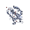
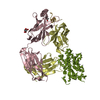
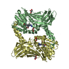
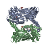
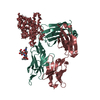
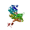
Crystal Structure of the ankyrin-B ZU5-ZU5-UPA-DD tandem
Components
Ankyrin-2
Keywords
PROTEIN BINDING / ZU5 / UPA / death domain / supramodule
Function / homology
Function and homology information
protein localization to T-tubule / atrial cardiac muscle cell to AV node cell communication / SA node cell to atrial cardiac muscle cell communication / protein localization to M-band / T-tubule organization / SA node cell action potential / membrane depolarization during SA node cell action potential / paranodal junction assembly / potassium channel activator activity / positive regulation of potassium ion import across plasma membrane ...protein localization to T-tubule / atrial cardiac muscle cell to AV node cell communication / SA node cell to atrial cardiac muscle cell communication / protein localization to M-band / T-tubule organization / SA node cell action potential / membrane depolarization during SA node cell action potential / paranodal junction assembly / potassium channel activator activity / positive regulation of potassium ion import across plasma membrane / regulation of atrial cardiac muscle cell action potential / channel activator activity / phosphorylation-dependent protein binding / sarcoplasmic reticulum calcium ion transport / regulation of SA node cell action potential / atrial cardiac muscle cell action potential / protein localization to endoplasmic reticulum / A band / atrial septum development / cytoskeletal adaptor activity / costamere / ventricular cardiac muscle cell action potential / regulation of ventricular cardiac muscle cell membrane repolarization / positive regulation of calcium ion transport / response to methylmercury / regulation of release of sequestered calcium ion into cytosol / regulation of cardiac muscle cell contraction / protein localization to cell surface / M band / regulation of cardiac muscle contraction by calcium ion signaling / Interaction between L1 and Ankyrins / spectrin binding / regulation of calcium ion transport / regulation of heart rate by cardiac conduction / intercalated disc / regulation of cardiac muscle contraction / COPI-mediated anterograde transport / regulation of cardiac muscle contraction by regulation of the release of sequestered calcium ion / T-tubule / regulation of heart rate / protein localization to plasma membrane / regulation of protein stability / sarcolemma / structural constituent of cytoskeleton / recycling endosome / Z disc / intracellular calcium ion homeostasis / endocytosis / intracellular protein localization / protein transport / ATPase binding / basolateral plasma membrane / cytoskeleton / transmembrane transporter binding / protein-macromolecule adaptor activity / early endosome / postsynaptic membrane / lysosome / apical plasma membrane / neuron projection / protein stabilization / positive regulation of gene expression / protein kinase binding / enzyme binding / mitochondrion / plasma membrane / cytosol Similarity search - Function
Immunoglobulin-like - #2660 / Chondroitinase Ac; Chain A, domain 3 - #30 / Ankyrin, UPA domain / UPA domain / : / Chondroitinase Ac; Chain A, domain 3 / Domain present in ZO-1 and Unc5-like netrin receptors / ZU5 domain / ZU5 domain / ZU5 domain profile. ...Immunoglobulin-like - #2660 / Chondroitinase Ac; Chain A, domain 3 - #30 / Ankyrin, UPA domain / UPA domain / : / Chondroitinase Ac; Chain A, domain 3 / Domain present in ZO-1 and Unc5-like netrin receptors / ZU5 domain / ZU5 domain / ZU5 domain profile. / Death Domain, Fas / Death Domain, Fas / Death domain profile. / DEATH domain, found in proteins involved in cell death (apoptosis). / Death domain / Death domain / Ankyrin repeats (many copies) / Death-like domain superfamily / Ankyrin repeat / Ankyrin repeat profile. / Ankyrin repeats (3 copies) / Ankyrin repeat region circular profile. / ankyrin repeats / Ankyrin repeat / Ankyrin repeat-containing domain superfamily / Immunoglobulin-like / Sandwich / Orthogonal Bundle / Mainly Beta / Mainly Alpha Similarity search - Domain/homology
Mass: 18.015 Da / Num. of mol.: 101 / Source method: isolated from a natural source / Formula: H2O
Sequence details
A PART OF RESIDUES 1211-1219 IN Q01484 HAS BEEN DELETED. THE SEQUENCE OF RESIDUES 1220-1535 IS FROM ...A PART OF RESIDUES 1211-1219 IN Q01484 HAS BEEN DELETED. THE SEQUENCE OF RESIDUES 1220-1535 IS FROM ISOFORM 2 OF ANKYRIN-2 (Q01484-2).
-
Experimental details
-
Experiment
Experiment
Method: X-RAY DIFFRACTION / Number of used crystals: 1
-
Sample preparation
Crystal
Density Matthews: 2.18 Å3/Da / Density % sol: 43.45 % / Mosaicity: 0.312 °
Crystal grow
Temperature: 289 K / Method: vapor diffusion, hanging drop / pH: 7.5 Details: 18% PEG3350, 0.2M ammonium acetate, 5% n-Octyl-beta-D-glucoside, pH 7.5, vapor diffusion, hanging drop, temperature 289K
In the structure databanks used in Yorodumi, some data are registered as the other names, "COVID-19 virus" and "2019-nCoV". Here are the details of the virus and the list of structure data.
Jan 31, 2019. EMDB accession codes are about to change! (news from PDBe EMDB page)
EMDB accession codes are about to change! (news from PDBe EMDB page)
The allocation of 4 digits for EMDB accession codes will soon come to an end. Whilst these codes will remain in use, new EMDB accession codes will include an additional digit and will expand incrementally as the available range of codes is exhausted. The current 4-digit format prefixed with “EMD-” (i.e. EMD-XXXX) will advance to a 5-digit format (i.e. EMD-XXXXX), and so on. It is currently estimated that the 4-digit codes will be depleted around Spring 2019, at which point the 5-digit format will come into force.
The EM Navigator/Yorodumi systems omit the EMD- prefix.
Related info.:Q: What is EMD? / ID/Accession-code notation in Yorodumi/EM Navigator
Yorodumi is a browser for structure data from EMDB, PDB, SASBDB, etc.
This page is also the successor to EM Navigator detail page, and also detail information page/front-end page for Omokage search.
The word "yorodu" (or yorozu) is an old Japanese word meaning "ten thousand". "mi" (miru) is to see.
Related info.:EMDB / PDB / SASBDB / Comparison of 3 databanks / Yorodumi Search / Aug 31, 2016. New EM Navigator & Yorodumi / Yorodumi Papers / Jmol/JSmol / Function and homology information / Changes in new EM Navigator and Yorodumi
 Movie
Movie Controller


 Open data
 Basic information
 Components
 Keywords
 Function and homology information
 Homo sapiens (human)
 X-RAY DIFFRACTION /
  Authors
 Citation
 Structure visualization
 Downloads & links
 Other downloads
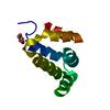


 PDBj
PDBj


 Assembly
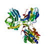

 Mass: 18.015 Da / Num. of mol.: 101 / Source method: isolated from a natural source / Formula: H2O
 Sample preparation
 / Beamline: BL17U / Wavelength: 0.98 Å
 Processing