 Movie
Movie Controller

[English] 日本語
 Yorodumi
- PDB-6m2e: Sirohydrochlorin nickelochelatase CfbA in complex with sirohydroc... -
+ Open data

- Basic information
| Entry | Database: PDB / ID: 6m2e | ||||||
|---|---|---|---|---|---|---|---|
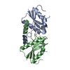
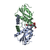
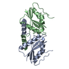
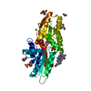
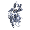
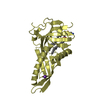
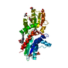
| Title | Sirohydrochlorin nickelochelatase CfbA in complex with sirohydrochlorin | ||||||
 Components Components | Sirohydrochlorin cobaltochelatase | ||||||
 Keywords Keywords | BIOSYNTHETIC PROTEIN / Chelatase | ||||||
| Function / homology |  Function and homology information Function and homology informationsirohydrochlorin nickelchelatase / sirohydrochlorin cobaltochelatase / sirohydrochlorin cobaltochelatase activity / : / methanogenesis / cobalt ion binding / nickel cation binding Similarity search - Function | ||||||
| Biological species |   Methanocaldococcus jannaschii (archaea) Methanocaldococcus jannaschii (archaea) | ||||||
| Method |  X-RAY DIFFRACTION / SYNCHROTRON / MOLECULAR REPLACEMENT / Resolution: 2.6 Å X-RAY DIFFRACTION / SYNCHROTRON / MOLECULAR REPLACEMENT / Resolution: 2.6 Å | ||||||
 Authors Authors | Fujishiro, T. | ||||||
| Funding support |  Japan, 1items Japan, 1items
| ||||||
 Citation Citation | Journal: Chem Sci / Year: 2021 Title: The nickel-sirohydrochlorin formation mechanism of the ancestral class II chelatase CfbA in coenzyme F430 biosynthesis. Authors: Fujishiro, T. / Ogawa, S. | ||||||
| History |
|
- Structure visualization
| Structure viewer | Molecule: MolmilJmol/JSmol |
|---|
- Downloads & links
-Download
| PDBx/mmCIF format | 6m2e.cif.gz | 64.1 KB | Display | PDBx/mmCIF format |
|---|---|---|---|---|
| PDB format | pdb6m2e.ent.gz | 45.1 KB | Display | PDB format |
| PDBx/mmJSON format | 6m2e.json.gz | Tree view | PDBx/mmJSON format | |
| Others |  Other downloads Other downloads |
-Validation report
| Arichive directory | https://data.pdbj.org/pub/pdb/validation_reports/m2/6m2eftp://data.pdbj.org/pub/pdb/validation_reports/m2/6m2e | HTTPS FTP |
|---|
-Related structure data
| Related structure data | 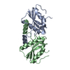 6m25C  6m26C 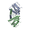 6m27C  6m28C 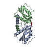 6m29C 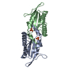 6m2aC 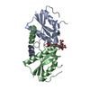 6m2fC 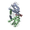 6m2gC  6m2hC  2xwsS S: Starting model for refinement C: citing same article ( |
|---|---|
| Similar structure data |


-Links
 PDBj
PDBj- Assembly
| Deposited unit | 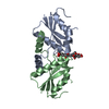
| ||||||||||||||||||
|---|---|---|---|---|---|---|---|---|---|---|---|---|---|---|---|---|---|---|---|
| 1 |
| ||||||||||||||||||
| Unit cell |
| ||||||||||||||||||
| Noncrystallographic symmetry (NCS) | NCS domain:
NCS domain segments: Component-ID: _ / Ens-ID: 1 / Beg auth comp-ID: MET / Beg label comp-ID: MET / End auth comp-ID: ARG / End label comp-ID: ARG / Refine code: _ / Auth seq-ID: 1 - 143 / Label seq-ID: 1 - 143
|
-Components
| #1: Protein | Mass: 16554.045 Da / Num. of mol.: 2 Source method: isolated from a genetically manipulated source Source: (gene. exp.) Methanocaldococcus jannaschii (strain ATCC 43067 / DSM 2661 / JAL-1 / JCM 10045 / NBRC 100440) (archaea)Gene: cbiX, cfbA, MJ0970 / Production host:  References: UniProt: Q58380, sirohydrochlorin cobaltochelatase, sirohydrochlorin nickelchelatase #2: Chemical | ChemComp-SHN / | 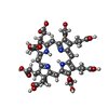  Mass: 862.832 Da / Num. of mol.: 1 / Source method: obtained synthetically / Formula: C42H46N4O16 / Feature type: SUBJECT OF INVESTIGATION Mass: 862.832 Da / Num. of mol.: 1 / Source method: obtained synthetically / Formula: C42H46N4O16 / Feature type: SUBJECT OF INVESTIGATION#3: Water | ChemComp-HOH / |  Mass: 18.015 Da / Num. of mol.: 5 / Source method: isolated from a natural source / Formula: H2O Mass: 18.015 Da / Num. of mol.: 5 / Source method: isolated from a natural source / Formula: H2OHas ligand of interest | Y | |
|---|
-Experimental details
-Experiment
| Experiment | Method: X-RAY DIFFRACTION / Number of used crystals: 1 |
|---|
- Sample preparation
| Crystal | Density Matthews: 2.95 Å3/Da / Density % sol: 58.27 % |
|---|---|
| Crystal grow | Temperature: 293 K / Method: vapor diffusion, sitting drop / pH: 5 Details: 0.1M ammonium sulfate, 0.3M Na-formate, 0.1M Na-acetate, 3% (w/v) gamma-PGA, 5% (w/v) PEG 3350 |
-Data collection
| Diffraction | Mean temperature: 100 K / Serial crystal experiment: N | ||||||||||||||||||||||||||||||||||||||||||||||||||||||||||||||||||||||||||||||||||||||||||||||||||||||||||||||||||||||||||||||||||||||||||||
|---|---|---|---|---|---|---|---|---|---|---|---|---|---|---|---|---|---|---|---|---|---|---|---|---|---|---|---|---|---|---|---|---|---|---|---|---|---|---|---|---|---|---|---|---|---|---|---|---|---|---|---|---|---|---|---|---|---|---|---|---|---|---|---|---|---|---|---|---|---|---|---|---|---|---|---|---|---|---|---|---|---|---|---|---|---|---|---|---|---|---|---|---|---|---|---|---|---|---|---|---|---|---|---|---|---|---|---|---|---|---|---|---|---|---|---|---|---|---|---|---|---|---|---|---|---|---|---|---|---|---|---|---|---|---|---|---|---|---|---|---|---|
| Diffraction source | Source: SYNCHROTRON / Site: SPring-8 / Beamline: BL44XU / Wavelength: 1.60699 Å | ||||||||||||||||||||||||||||||||||||||||||||||||||||||||||||||||||||||||||||||||||||||||||||||||||||||||||||||||||||||||||||||||||||||||||||
| Detector | Type: DECTRIS EIGER X 16M / Detector: PIXEL / Date: Jul 22, 2019 | ||||||||||||||||||||||||||||||||||||||||||||||||||||||||||||||||||||||||||||||||||||||||||||||||||||||||||||||||||||||||||||||||||||||||||||
| Radiation | Monochromator: liquid-nitrogen-cooled Si(111) double-crystal monochromator Protocol: SINGLE WAVELENGTH / Monochromatic (M) / Laue (L): M / Scattering type: x-ray | ||||||||||||||||||||||||||||||||||||||||||||||||||||||||||||||||||||||||||||||||||||||||||||||||||||||||||||||||||||||||||||||||||||||||||||
| Radiation wavelength | Wavelength: 1.60699 Å / Relative weight: 1 | ||||||||||||||||||||||||||||||||||||||||||||||||||||||||||||||||||||||||||||||||||||||||||||||||||||||||||||||||||||||||||||||||||||||||||||
| Reflection | Resolution: 2.6→48.89 Å / Num. obs: 11899 / % possible obs: 100 % / Redundancy: 13.665 % / Biso Wilson estimate: 83.4 Å2 / CC1/2: 0.999 / Rmerge(I) obs: 0.059 / Rrim(I) all: 0.061 / Χ2: 0.97 / Net I/σ(I): 25.66 / Num. measured all: 162601 / Scaling rejects: 13 | ||||||||||||||||||||||||||||||||||||||||||||||||||||||||||||||||||||||||||||||||||||||||||||||||||||||||||||||||||||||||||||||||||||||||||||
| Reflection shell | Diffraction-ID: 1
|
- Processing
| Software |
| |||||||||||||||||||||||||||||||||||||||||||||
|---|---|---|---|---|---|---|---|---|---|---|---|---|---|---|---|---|---|---|---|---|---|---|---|---|---|---|---|---|---|---|---|---|---|---|---|---|---|---|---|---|---|---|---|---|---|---|
| Refinement | Method to determine structure: MOLECULAR REPLACEMENT Starting model: 2XWS Resolution: 2.6→48.89 Å / Cor.coef. Fo:Fc: 0.963 / Cor.coef. Fo:Fc free: 0.942 / WRfactor Rfree: 0.253 / WRfactor Rwork: 0.2019 / FOM work R set: 0.7885 / SU B: 10.01 / SU ML: 0.206 / SU R Cruickshank DPI: 0.3808 / SU Rfree: 0.2581 / Cross valid method: THROUGHOUT / σ(F): 0 / ESU R: 0.381 / ESU R Free: 0.258 / Stereochemistry target values: MAXIMUM LIKELIHOOD / Details: U VALUES : REFINED INDIVIDUALLY
| |||||||||||||||||||||||||||||||||||||||||||||
| Solvent computation | Ion probe radii: 0.8 Å / Shrinkage radii: 0.8 Å / VDW probe radii: 1.2 Å / Solvent model: MASK | |||||||||||||||||||||||||||||||||||||||||||||
| Displacement parameters | Biso max: 175.45 Å2 / Biso mean: 82.477 Å2 / Biso min: 44.27 Å2
| |||||||||||||||||||||||||||||||||||||||||||||
| Refinement step | Cycle: final / Resolution: 2.6→48.89 Å
| |||||||||||||||||||||||||||||||||||||||||||||
| Refine LS restraints |
| |||||||||||||||||||||||||||||||||||||||||||||
| Refine LS restraints NCS | Ens-ID: 1 / Number: 3568 / Refine-ID: X-RAY DIFFRACTION / Type: interatomic distance / Rms dev position: 0.08 Å / Weight position: 0.05
| |||||||||||||||||||||||||||||||||||||||||||||
| LS refinement shell | Resolution: 2.6→2.667 Å / Rfactor Rfree error: 0 / Total num. of bins used: 20
|
