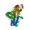
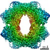
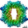
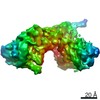
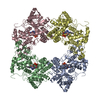
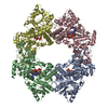
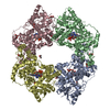
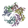
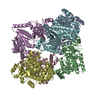
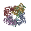
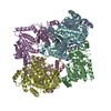
Journal: Nat Struct Mol Biol / Year: 2020 Title: The oligomeric structures of plant cryptochromes. Authors: Kai Shao / Xue Zhang / Xu Li / Yahui Hao / Xiaowei Huang / Miaolian Ma / Minhua Zhang / Fang Yu / Hongtao Liu / Peng Zhang / Abstract: Cryptochromes (CRYs) are a group of evolutionarily conserved flavoproteins found in many organisms. In plants, the well-studied CRY photoreceptor, activated by blue light, plays essential roles in ...Cryptochromes (CRYs) are a group of evolutionarily conserved flavoproteins found in many organisms. In plants, the well-studied CRY photoreceptor, activated by blue light, plays essential roles in plant growth and development. However, the mechanism of activation remains largely unknown. Here, we determined the oligomeric structures of the blue-light-perceiving PHR domain of Zea mays CRY1 and an Arabidopsis CRY2 constitutively active mutant. The structures form dimers and tetramers whose functional importance is examined in vitro and in vivo with Arabidopsis CRY2. Structure-based analysis suggests that blue light may be perceived by CRY to cause conformational changes, whose precise nature remains to be determined, leading to oligomerization that is essential for downstream signaling. This photoactivation mechanism may be widely used by plant CRYs. Our study reveals a molecular mechanism of plant CRY activation and also paves the way for design of CRY as a more efficient optical switch.
In the structure databanks used in Yorodumi, some data are registered as the other names, "COVID-19 virus" and "2019-nCoV". Here are the details of the virus and the list of structure data.
Jan 31, 2019. EMDB accession codes are about to change! (news from PDBe EMDB page)
EMDB accession codes are about to change! (news from PDBe EMDB page)
The allocation of 4 digits for EMDB accession codes will soon come to an end. Whilst these codes will remain in use, new EMDB accession codes will include an additional digit and will expand incrementally as the available range of codes is exhausted. The current 4-digit format prefixed with “EMD-” (i.e. EMD-XXXX) will advance to a 5-digit format (i.e. EMD-XXXXX), and so on. It is currently estimated that the 4-digit codes will be depleted around Spring 2019, at which point the 5-digit format will come into force.
The EM Navigator/Yorodumi systems omit the EMD- prefix.
Related info.:Q: What is EMD? / ID/Accession-code notation in Yorodumi/EM Navigator
Yorodumi is a browser for structure data from EMDB, PDB, SASBDB, etc.
This page is also the successor to EM Navigator detail page, and also detail information page/front-end page for Omokage search.
The word "yorodu" (or yorozu) is an old Japanese word meaning "ten thousand". "mi" (miru) is to see.
Related info.:EMDB / PDB / SASBDB / Comparison of 3 databanks / Yorodumi Search / Aug 31, 2016. New EM Navigator & Yorodumi / Yorodumi Papers / Jmol/JSmol / Function and homology information / Changes in new EM Navigator and Yorodumi
 Movie
Movie Controller


 Open data
 Basic information
 Components
 Keywords
 Function and homology information
 X-RAY DIFFRACTION /
  Authors
 Citation
 Structure visualization
 Downloads & links
 Other downloads


 PDBj
PDBj
 Assembly
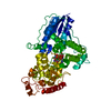


 Spodoptera frugiperda (fall armyworm) / References: UniProt: A0A1D6HB66, UniProt: A0A1D6HB67*PLUS
 Mass: 785.550 Da / Num. of mol.: 1 / Source method: obtained synthetically / Formula: C27H33N9O15P2 / Feature type: SUBJECT OF INVESTIGATION / Comment: FAD*YM
 Sample preparation
 Processing