flavin adenine dinucleotide metabolic process / regulation of meristem growth / long-day photoperiodism, flowering / response to absence of light / circadian regulation of calcium ion oscillation / response to low fluence blue light stimulus by blue low-fluence system / regulation of flower development / positive regulation of flower development / response to strigolactone / regulation of leaf morphogenesis ...flavin adenine dinucleotide metabolic process / regulation of meristem growth / long-day photoperiodism, flowering / response to absence of light / circadian regulation of calcium ion oscillation / response to low fluence blue light stimulus by blue low-fluence system / regulation of flower development / positive regulation of flower development / response to strigolactone / regulation of leaf morphogenesis / regulation of photoperiodism, flowering / phototropism / blue light signaling pathway / stomatal movement / deoxyribodipyrimidine photo-lyase activity / blue light photoreceptor activity / response to blue light / response to water deprivation / plant-type vacuole / entrainment of circadian clock by photoperiod / response to light stimulus / FAD binding / circadian regulation of gene expression / circadian rhythm / regulation of circadian rhythm / positive regulation of reactive oxygen species metabolic process / chromatin organization / defense response to virus / nuclear body / chromatin remodeling / protein homodimerization activity / DNA binding / ATP binding / metal ion binding / identical protein binding / nucleus / cytoplasm Similarity search - Function
Cryptochrome, plant / DNA photolyases class 1 signature 2. / Cryptochrome/DNA photolyase class 1, conserved site, C-terminal / DNA photolyases class 1 signature 1. / Cryptochrome/DNA photolyase class 1 / Cryptochrome/DNA photolyase, FAD-binding domain / FAD binding domain of DNA photolyase / DNA photolyase, N-terminal / Cryptochrome/photolyase, N-terminal domain superfamily / DNA photolyase ...Cryptochrome, plant / DNA photolyases class 1 signature 2. / Cryptochrome/DNA photolyase class 1, conserved site, C-terminal / DNA photolyases class 1 signature 1. / Cryptochrome/DNA photolyase class 1 / Cryptochrome/DNA photolyase, FAD-binding domain / FAD binding domain of DNA photolyase / DNA photolyase, N-terminal / Cryptochrome/photolyase, N-terminal domain superfamily / DNA photolyase / Photolyase/cryptochrome alpha/beta domain profile. / Cryptochrome/DNA photolyase, FAD-binding domain-like superfamily / Rossmann-like alpha/beta/alpha sandwich fold Similarity search - Domain/homology
In the structure databanks used in Yorodumi, some data are registered as the other names, "COVID-19 virus" and "2019-nCoV". Here are the details of the virus and the list of structure data.
Jan 31, 2019. EMDB accession codes are about to change! (news from PDBe EMDB page)
EMDB accession codes are about to change! (news from PDBe EMDB page)
The allocation of 4 digits for EMDB accession codes will soon come to an end. Whilst these codes will remain in use, new EMDB accession codes will include an additional digit and will expand incrementally as the available range of codes is exhausted. The current 4-digit format prefixed with “EMD-” (i.e. EMD-XXXX) will advance to a 5-digit format (i.e. EMD-XXXXX), and so on. It is currently estimated that the 4-digit codes will be depleted around Spring 2019, at which point the 5-digit format will come into force.
The EM Navigator/Yorodumi systems omit the EMD- prefix.
Related info.:Q: What is EMD? / ID/Accession-code notation in Yorodumi/EM Navigator
Yorodumi is a browser for structure data from EMDB, PDB, SASBDB, etc.
This page is also the successor to EM Navigator detail page, and also detail information page/front-end page for Omokage search.
The word "yorodu" (or yorozu) is an old Japanese word meaning "ten thousand". "mi" (miru) is to see.
Related info.:EMDB / PDB / SASBDB / Comparison of 3 databanks / Yorodumi Search / Aug 31, 2016. New EM Navigator & Yorodumi / Yorodumi Papers / Jmol/JSmol / Function and homology information / Changes in new EM Navigator and Yorodumi
 Movie
Movie Controller


 Open data
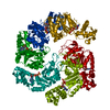
 Basic information
 Components
 Keywords
 Function and homology information
 X-RAY DIFFRACTION /
  Authors
 Citation
 Structure visualization
 Downloads & links
 Other downloads
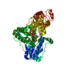

 PDBj
PDBj
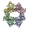
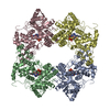
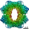
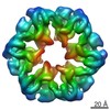
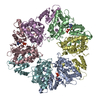
 Assembly
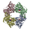
 Baculovirus expression vector pFastBac1-HM / References: UniProt: Q96524
 Mass: 785.550 Da / Num. of mol.: 4 / Source method: obtained synthetically / Formula: C27H33N9O15P2 / Feature type: SUBJECT OF INVESTIGATION / Comment: FAD*YM
 Mass: 92.094 Da / Num. of mol.: 15 / Source method: obtained synthetically / Formula: C3H8O3
 Mass: 18.015 Da / Num. of mol.: 30 / Source method: isolated from a natural source / Formula: H2O
 Sample preparation
 / Beamline: 8.2.1 / Wavelength: 1 Å
 Processing