 Movie
Movie Controller


+ Open data

- Basic information
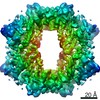
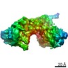
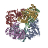
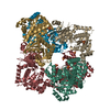
| Entry | Database: PDB / ID: 6lz3 | |||||||||||||||||||||||||||||||||||||||
|---|---|---|---|---|---|---|---|---|---|---|---|---|---|---|---|---|---|---|---|---|---|---|---|---|---|---|---|---|---|---|---|---|---|---|---|---|---|---|---|---|
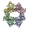
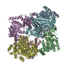
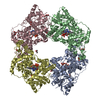
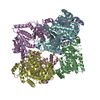
| Title | Structure of cryptochrome in active conformation | |||||||||||||||||||||||||||||||||||||||
 Components Components | Cryptochrome2 | |||||||||||||||||||||||||||||||||||||||
 Keywords Keywords | FLAVOPROTEIN / cryptochrome / photoreceptor / photosignaling / PLANT PROTEIN | |||||||||||||||||||||||||||||||||||||||
| Function / homology |  Function and homology information Function and homology informationblue light photoreceptor activity / nucleobase-containing compound metabolic process / response to stress / nucleotide binding Similarity search - Function | |||||||||||||||||||||||||||||||||||||||
| Biological species |  | |||||||||||||||||||||||||||||||||||||||
| Method | ELECTRON MICROSCOPY / single particle reconstruction / cryo EM / Resolution: 3.2 Å | |||||||||||||||||||||||||||||||||||||||
 Authors Authors | Shao, K. / Zhang, X. / Zhang, P. | |||||||||||||||||||||||||||||||||||||||
 Citation Citation | Journal: Nat Struct Mol Biol / Year: 2020 Title: The oligomeric structures of plant cryptochromes. Authors: Kai Shao / Xue Zhang / Xu Li / Yahui Hao / Xiaowei Huang / Miaolian Ma / Minhua Zhang / Fang Yu / Hongtao Liu / Peng Zhang /  Abstract: Cryptochromes (CRYs) are a group of evolutionarily conserved flavoproteins found in many organisms. In plants, the well-studied CRY photoreceptor, activated by blue light, plays essential roles in ...Cryptochromes (CRYs) are a group of evolutionarily conserved flavoproteins found in many organisms. In plants, the well-studied CRY photoreceptor, activated by blue light, plays essential roles in plant growth and development. However, the mechanism of activation remains largely unknown. Here, we determined the oligomeric structures of the blue-light-perceiving PHR domain of Zea mays CRY1 and an Arabidopsis CRY2 constitutively active mutant. The structures form dimers and tetramers whose functional importance is examined in vitro and in vivo with Arabidopsis CRY2. Structure-based analysis suggests that blue light may be perceived by CRY to cause conformational changes, whose precise nature remains to be determined, leading to oligomerization that is essential for downstream signaling. This photoactivation mechanism may be widely used by plant CRYs. Our study reveals a molecular mechanism of plant CRY activation and also paves the way for design of CRY as a more efficient optical switch. | |||||||||||||||||||||||||||||||||||||||
| History |
|
- Structure visualization
| Movie |
Movie viewer |
|---|---|
| Structure viewer | Molecule: MolmilJmol/JSmol |
- Downloads & links
-Download
| PDBx/mmCIF format | 6lz3.cif.gz | 359.2 KB | Display | PDBx/mmCIF format |
|---|---|---|---|---|
| PDB format | pdb6lz3.ent.gz | 292.1 KB | Display | PDB format |
| PDBx/mmJSON format | 6lz3.json.gz | Tree view | PDBx/mmJSON format | |
| Others |  Other downloads Other downloads |
-Validation report
| Arichive directory | https://data.pdbj.org/pub/pdb/validation_reports/lz/6lz3ftp://data.pdbj.org/pub/pdb/validation_reports/lz/6lz3 | HTTPS FTP |
|---|
-Related structure data
| Related structure data | 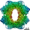 30022MC 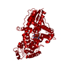 6lz7C M: map data used to model this data C: citing same article ( |
|---|---|
| Similar structure data |


-Links
 PDBj
PDBj
- Assembly
| Deposited unit | 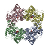
|
|---|---|
| 1 |
|
-Components
| #1: Protein | Mass: 77609.125 Da / Num. of mol.: 4 / Mutation: W368A Source method: isolated from a genetically manipulated source Source: (gene. exp.)   Spodoptera frugiperda (fall armyworm) / References: UniProt: B8A2L5 Spodoptera frugiperda (fall armyworm) / References: UniProt: B8A2L5#2: Chemical | ChemComp-FAD /   Mass: 785.550 Da / Num. of mol.: 4 / Source method: obtained synthetically / Formula: C27H33N9O15P2 / Feature type: SUBJECT OF INVESTIGATION / Comment: FAD*YM Mass: 785.550 Da / Num. of mol.: 4 / Source method: obtained synthetically / Formula: C27H33N9O15P2 / Feature type: SUBJECT OF INVESTIGATION / Comment: FAD*YMHas ligand of interest | Y | Has protein modification | N | |
|---|
-Experimental details
-Experiment
| Experiment | Method: ELECTRON MICROSCOPY |
|---|---|
| EM experiment | Aggregation state: PARTICLE / 3D reconstruction method: single particle reconstruction |
- Sample preparation
| Component | Name: Tetrameric complex of ZmCRY1c / Type: COMPLEX / Entity ID: #1 / Source: RECOMBINANT |
|---|---|
| Source (natural) | Organism: |
| Source (recombinant) | Organism: Spodoptera frugiperda (fall armyworm) |
| Buffer solution | pH: 8 |
| Specimen | Embedding applied: NO / Shadowing applied: NO / Staining applied: NO / Vitrification applied: YES |
| Vitrification | Cryogen name: ETHANE |
- Electron microscopy imaging
| Experimental equipment |  Model: Titan Krios / Image courtesy: FEI Company |
|---|---|
| Microscopy | Model: FEI TITAN KRIOS |
| Electron gun | Electron source:  FIELD EMISSION GUN / Accelerating voltage: 300 kV / Illumination mode: OTHER FIELD EMISSION GUN / Accelerating voltage: 300 kV / Illumination mode: OTHER |
| Electron lens | Mode: OTHER |
| Image recording | Electron dose: 49.8 e/Å2 / Film or detector model: GATAN K2 SUMMIT (4k x 4k) |
- Processing
| CTF correction | Type: NONE |
|---|---|
| Symmetry | Point symmetry: D2 (2x2 fold dihedral) |
| 3D reconstruction | Resolution: 3.2 Å / Resolution method: FSC 0.143 CUT-OFF / Num. of particles: 108059 / Symmetry type: POINT |
