 Movie
Movie Controller

[English] 日本語
 Yorodumi
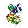
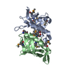
- PDB-6lwu: Complex of Gynuella sunshinyii GH46 chitosanase GsCsn46A E19A wit... -
+ Open data

- Basic information
| Entry | Database: PDB / ID: 6lwu | ||||||
|---|---|---|---|---|---|---|---|
| Title | Complex of Gynuella sunshinyii GH46 chitosanase GsCsn46A E19A with chitopentaose | ||||||
 Components Components | chitosanase | ||||||
 Keywords Keywords | HYDROLASE / chitosanase / glucoside hydrolase / GH46 | ||||||
| Function / homology | Glycoside hydrolase, family 46, N-terminal / Glycosyl hydrolase family 46 / chitosanase activity / Glycoside hydrolase, family 46 / Lysozyme-like domain superfamily / carbohydrate metabolic process / extracellular region / 2-amino-2-deoxy-beta-D-glucopyranose / Chitosanase Function and homology information Function and homology information | ||||||
| Biological species |  Gynuella sunshinyii YC6258 (bacteria) Gynuella sunshinyii YC6258 (bacteria) | ||||||
| Method |  X-RAY DIFFRACTION / SYNCHROTRON / MOLECULAR REPLACEMENT / Resolution: 2.419 Å X-RAY DIFFRACTION / SYNCHROTRON / MOLECULAR REPLACEMENT / Resolution: 2.419 Å | ||||||
 Authors Authors | Qin, Z. / Wang, Y. | ||||||
| Funding support |  China, 1items China, 1items
| ||||||
 Citation Citation | Journal: To Be Published Title: Complex of Gynuella sunshinyii GH46 chitosanase GsCsn46A E19A with chitopentaose Authors: Qin, Z. / Wang, Y. | ||||||
| History |
|
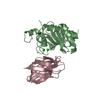
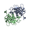
- Structure visualization
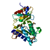
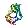
| Structure viewer | Molecule: MolmilJmol/JSmol |
|---|
- Downloads & links
-Download
| PDBx/mmCIF format | 6lwu.cif.gz | 63.9 KB | Display | PDBx/mmCIF format |
|---|---|---|---|---|
| PDB format | pdb6lwu.ent.gz | 44.8 KB | Display | PDB format |
| PDBx/mmJSON format | 6lwu.json.gz | Tree view | PDBx/mmJSON format | |
| Others |  Other downloads Other downloads |
-Validation report
| Arichive directory | https://data.pdbj.org/pub/pdb/validation_reports/lw/6lwuftp://data.pdbj.org/pub/pdb/validation_reports/lw/6lwu | HTTPS FTP |
|---|
-Related structure data
| Related structure data | 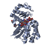 4oltS S: Starting model for refinement |
|---|---|
| Similar structure data |


-Links
 PDBj
PDBj- Assembly
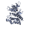
| Deposited unit | 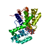
| ||||||||
|---|---|---|---|---|---|---|---|---|---|
| 1 |
| ||||||||
| Unit cell |
|
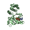
-Components
| #1: Protein | Mass: 27057.549 Da / Num. of mol.: 1 / Mutation: E42A Source method: isolated from a genetically manipulated source Source: (gene. exp.) Gynuella sunshinyii YC6258 (bacteria) / Gene: YC6258_05941Production host: References: UniProt: A0A0C5VFC0 | ||||
|---|---|---|---|---|---|
| #2: Polysaccharide | 2-amino-2-deoxy-beta-D-glucopyranose-(1-4)-2-amino-2-deoxy-beta-D-glucopyranose-(1-4)-2-amino-2- ...2-amino-2-deoxy-beta-D-glucopyranose-(1-4)-2-amino-2-deoxy-beta-D-glucopyranose-(1-4)-2-amino-2-deoxy-beta-D-glucopyranose Source method: isolated from a genetically manipulated source | ||||
| #3: Sugar | 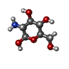  Type: D-saccharide, beta linking / Mass: 179.171 Da / Num. of mol.: 2 Type: D-saccharide, beta linking / Mass: 179.171 Da / Num. of mol.: 2Source method: isolated from a genetically manipulated source Formula: C6H13NO5 / Feature type: SUBJECT OF INVESTIGATION #4: Water | ChemComp-HOH / |  Mass: 18.015 Da / Num. of mol.: 52 / Source method: isolated from a natural source / Formula: H2O Mass: 18.015 Da / Num. of mol.: 52 / Source method: isolated from a natural source / Formula: H2OHas ligand of interest | Y | |
-Experimental details
-Experiment
| Experiment | Method: X-RAY DIFFRACTION / Number of used crystals: 1 |
|---|
- Sample preparation
| Crystal | Density Matthews: 2.95 Å3/Da / Density % sol: 58.31 % |
|---|---|
| Crystal grow | Temperature: 293 K / Method: vapor diffusion, sitting drop / pH: 7.5 Details: 0.1M HEPES, 10%PEG 8000, 8% (v/v) glycol,2% (w/v) chitopentaose |
-Data collection
| Diffraction | Mean temperature: 100 K / Serial crystal experiment: N | |||||||||||||||||||||||||||||||||||||||||||||||||||||||||||||||||||||||||||||||||||||||||||||||||||||||||||||||||||||||||||||||||||||||||||||||||||||||||||||||||||||||||||||||||||||||||||||
|---|---|---|---|---|---|---|---|---|---|---|---|---|---|---|---|---|---|---|---|---|---|---|---|---|---|---|---|---|---|---|---|---|---|---|---|---|---|---|---|---|---|---|---|---|---|---|---|---|---|---|---|---|---|---|---|---|---|---|---|---|---|---|---|---|---|---|---|---|---|---|---|---|---|---|---|---|---|---|---|---|---|---|---|---|---|---|---|---|---|---|---|---|---|---|---|---|---|---|---|---|---|---|---|---|---|---|---|---|---|---|---|---|---|---|---|---|---|---|---|---|---|---|---|---|---|---|---|---|---|---|---|---|---|---|---|---|---|---|---|---|---|---|---|---|---|---|---|---|---|---|---|---|---|---|---|---|---|---|---|---|---|---|---|---|---|---|---|---|---|---|---|---|---|---|---|---|---|---|---|---|---|---|---|---|---|---|---|---|---|---|
| Diffraction source | Source: SYNCHROTRON / Site: APS  / Beamline: 17-BM / Wavelength: 0.9792 Å / Beamline: 17-BM / Wavelength: 0.9792 Å | |||||||||||||||||||||||||||||||||||||||||||||||||||||||||||||||||||||||||||||||||||||||||||||||||||||||||||||||||||||||||||||||||||||||||||||||||||||||||||||||||||||||||||||||||||||||||||||
| Detector | Type: MAR CCD 165 mm / Detector: CCD / Date: Dec 8, 2018 | |||||||||||||||||||||||||||||||||||||||||||||||||||||||||||||||||||||||||||||||||||||||||||||||||||||||||||||||||||||||||||||||||||||||||||||||||||||||||||||||||||||||||||||||||||||||||||||
| Radiation | Protocol: SINGLE WAVELENGTH / Monochromatic (M) / Laue (L): M / Scattering type: x-ray | |||||||||||||||||||||||||||||||||||||||||||||||||||||||||||||||||||||||||||||||||||||||||||||||||||||||||||||||||||||||||||||||||||||||||||||||||||||||||||||||||||||||||||||||||||||||||||||
| Radiation wavelength | Wavelength: 0.9792 Å / Relative weight: 1 | |||||||||||||||||||||||||||||||||||||||||||||||||||||||||||||||||||||||||||||||||||||||||||||||||||||||||||||||||||||||||||||||||||||||||||||||||||||||||||||||||||||||||||||||||||||||||||||
| Reflection | Resolution: 2.419→50 Å / Num. obs: 11676 / % possible obs: 99.2 % / Redundancy: 9.7 % / Rmerge(I) obs: 0.043 / Rpim(I) all: 0.015 / Rrim(I) all: 0.046 / Χ2: 0.811 / Net I/σ(I): 12.1 | |||||||||||||||||||||||||||||||||||||||||||||||||||||||||||||||||||||||||||||||||||||||||||||||||||||||||||||||||||||||||||||||||||||||||||||||||||||||||||||||||||||||||||||||||||||||||||||
| Reflection shell | Diffraction-ID: 1
|
- Processing
| Software |
| ||||||||||||||||||||||||||||||||||||||||||||||||||||||
|---|---|---|---|---|---|---|---|---|---|---|---|---|---|---|---|---|---|---|---|---|---|---|---|---|---|---|---|---|---|---|---|---|---|---|---|---|---|---|---|---|---|---|---|---|---|---|---|---|---|---|---|---|---|---|---|
| Refinement | Method to determine structure: MOLECULAR REPLACEMENT Starting model: 4OLT Resolution: 2.419→45.81 Å / SU ML: 0.28 / Cross valid method: THROUGHOUT / σ(F): 2.05 / Phase error: 30.18
| ||||||||||||||||||||||||||||||||||||||||||||||||||||||
| Solvent computation | Shrinkage radii: 0.9 Å / VDW probe radii: 1.11 Å | ||||||||||||||||||||||||||||||||||||||||||||||||||||||
| Displacement parameters | Biso max: 79.43 Å2 / Biso mean: 35.8523 Å2 / Biso min: 17.33 Å2 | ||||||||||||||||||||||||||||||||||||||||||||||||||||||
| Refinement step | Cycle: final / Resolution: 2.419→45.81 Å
| ||||||||||||||||||||||||||||||||||||||||||||||||||||||
| LS refinement shell | Refine-ID: X-RAY DIFFRACTION / Rfactor Rfree error: 0
|
