 Movie
Movie Controller

[English] 日本語
 Yorodumi
- PDB-5ai1: Crystal structure of ketosteroid isomerase containing Y32F, D40N,... -
+ Open data

- Basic information
| Entry | Database: PDB / ID: 5ai1 | ||||||
|---|---|---|---|---|---|---|---|
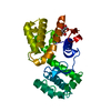
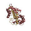
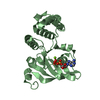
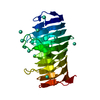
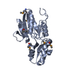
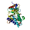
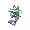
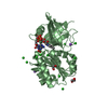
| Title | Crystal structure of ketosteroid isomerase containing Y32F, D40N, Y57F and Y119F mutations in the equilenin-bound form | ||||||
 Components Components | KETOSTEROID ISOMERASE | ||||||
 Keywords Keywords | ISOMERASE / KETOSTEROID ISOMERASE / PSEUDOMONAS PUTIDA / EQUILENIN | ||||||
| Function / homology |  Function and homology information Function and homology informationsteroid Delta-isomerase / steroid Delta-isomerase activity / steroid metabolic process Similarity search - Function | ||||||
| Biological species |  PSEUDOMONAS PUTIDA (bacteria) PSEUDOMONAS PUTIDA (bacteria) | ||||||
| Method |  X-RAY DIFFRACTION / SYNCHROTRON / MOLECULAR REPLACEMENT / Resolution: 2.103 Å X-RAY DIFFRACTION / SYNCHROTRON / MOLECULAR REPLACEMENT / Resolution: 2.103 Å | ||||||
 Authors Authors | Cha, H.J. / Jeong, J.H. / Kim, Y.G. | ||||||
 Citation Citation | Journal: Mol.Cells / Year: 2015 Title: Contribution of a Low-Barrier Hydrogen Bond to Catalysis is not Significant in Ketosteroid Isomerase. Authors: Jang, D.S. / Choi, G. / Cha, H.J. / Shin, S. / Hong, B.H. / Lee, H.J. / Lee, H.C. / Choi, K.Y. | ||||||
| History |
|
- Structure visualization
| Structure viewer | Molecule: MolmilJmol/JSmol |
|---|
- Downloads & links
-Download
| PDBx/mmCIF format | 5ai1.cif.gz | 63.6 KB | Display | PDBx/mmCIF format |
|---|---|---|---|---|
| PDB format | pdb5ai1.ent.gz | 46.6 KB | Display | PDB format |
| PDBx/mmJSON format | 5ai1.json.gz | Tree view | PDBx/mmJSON format | |
| Others |  Other downloads Other downloads |
-Validation report
| Arichive directory | https://data.pdbj.org/pub/pdb/validation_reports/ai/5ai1ftp://data.pdbj.org/pub/pdb/validation_reports/ai/5ai1 | HTTPS FTP |
|---|
-Related structure data
| Related structure data |  1opyS S: Starting model for refinement |
|---|---|
| Similar structure data |


-Links
 PDBj
PDBj
- Assembly
| Deposited unit | 
| ||||||||
|---|---|---|---|---|---|---|---|---|---|
| 1 | 
| ||||||||
| Unit cell |
|
-Components
| #1: Protein | Mass: 14499.515 Da / Num. of mol.: 1 / Mutation: YES Source method: isolated from a genetically manipulated source Source: (gene. exp.) PSEUDOMONAS PUTIDA (bacteria) / Variant: BIOTYPE B / Plasmid: PKK223-3 / Production host: |
|---|---|
| #2: Chemical | ChemComp-EQU /   Mass: 266.334 Da / Num. of mol.: 1 / Source method: obtained synthetically / Formula: C18H18O2 Mass: 266.334 Da / Num. of mol.: 1 / Source method: obtained synthetically / Formula: C18H18O2 |
| #3: Water | ChemComp-HOH /  Mass: 18.015 Da / Num. of mol.: 58 / Source method: isolated from a natural source / Formula: H2O Mass: 18.015 Da / Num. of mol.: 58 / Source method: isolated from a natural source / Formula: H2O |
-Experimental details
-Experiment
| Experiment | Method: X-RAY DIFFRACTION / Number of used crystals: 2 |
|---|
- Sample preparation
| Crystal | Density Matthews: 2.13 Å3/Da / Density % sol: 42.17 % / Description: NONE |
|---|---|
| Crystal grow | pH: 4.6 Details: 0.1M SODIUM ACETATE (PH 4.5), 0.6M AMMONIUM ACETATE, 30% PEG 4,000 |
-Data collection
| Diffraction | Mean temperature: 100 K |
|---|---|
| Diffraction source | Source: SYNCHROTRON / Site: PAL/PLS  / Beamline: 5C (4A) / Wavelength: 0.9796 / Beamline: 5C (4A) / Wavelength: 0.9796 |
| Detector | Type: ADSC QUANTUM 315r / Detector: CCD / Date: Dec 16, 2014 / Details: MIRRORS |
| Radiation | Monochromator: DOUBLE CRYSTAL MONOCHROMATOR / Protocol: SINGLE WAVELENGTH / Monochromatic (M) / Laue (L): M / Scattering type: x-ray |
| Radiation wavelength | Wavelength: 0.9796 Å / Relative weight: 1 |
| Reflection | Resolution: 2.1→30.13 Å / Num. obs: 7126 / % possible obs: 94.4 % / Observed criterion σ(I): 0 / Redundancy: 7.6 % / Biso Wilson estimate: 26.51 Å2 / Rmerge(I) obs: 0.08 / Net I/σ(I): 23 |
| Reflection shell | Resolution: 2.1→2.14 Å / Redundancy: 4.1 % / Rmerge(I) obs: 0.34 / Mean I/σ(I) obs: 2.9 / % possible all: 89.6 |
- Processing
| Software |
| ||||||||||||||||||||||||||||||||||||||||||
|---|---|---|---|---|---|---|---|---|---|---|---|---|---|---|---|---|---|---|---|---|---|---|---|---|---|---|---|---|---|---|---|---|---|---|---|---|---|---|---|---|---|---|---|
| Refinement | Method to determine structure: MOLECULAR REPLACEMENT Starting model: PDB ENTRY 1OPY Resolution: 2.103→30.134 Å / SU ML: 0.26 / σ(F): 1.5 / Phase error: 26.71 / Stereochemistry target values: ML
| ||||||||||||||||||||||||||||||||||||||||||
| Solvent computation | Shrinkage radii: 0.9 Å / VDW probe radii: 1.11 Å / Solvent model: FLAT BULK SOLVENT MODEL | ||||||||||||||||||||||||||||||||||||||||||
| Refinement step | Cycle: LAST / Resolution: 2.103→30.134 Å
| ||||||||||||||||||||||||||||||||||||||||||
| Refine LS restraints |
| ||||||||||||||||||||||||||||||||||||||||||
| LS refinement shell |
|
