HETEROGEN THE COORDINATES CONTAIN THE MINOR CONFORMATION OF OG SER 213. THE SHORT CONTACT BETWEEN ...HETEROGEN THE COORDINATES CONTAIN THE MINOR CONFORMATION OF OG SER 213. THE SHORT CONTACT BETWEEN THIS ATOM AND O2 SO4 301 PROBABLY INDICATES THAT THE SULFATE ION POSITION IS PARTIALLY OCCUPIED BY A WATER MOLECULE.
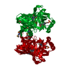
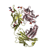
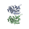
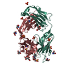
The second molecule of the dimer can be generated by applying the matrix -0.50000 -0.86603 0.00000 -0.86603 0.50000 -0.00000 0.00000 -0.00000 -1.00000 and the vector 84.33016 48.68816 103.37299
-
Components
#1: Protein
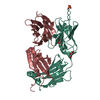
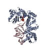
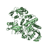
PhenazinebiosynthesisproteinphzF
Mass: 30085.191 Da / Num. of mol.: 1 Source method: isolated from a genetically manipulated source Source: (gene. exp.) Pseudomonas fluorescens (bacteria) / Gene: PHZF / Plasmid: pET28a / Species (production host): Escherichia coli / Production host: Escherichia coli BL21(DE3) (bacteria) / Strain (production host): BL21(DE3) / References: UniProt: Q51792
Mass: 18.015 Da / Num. of mol.: 253 / Source method: isolated from a natural source / Formula: H2O
Has protein modification
N
-
Experimental details
-
Experiment
Experiment
Method: X-RAY DIFFRACTION / Number of used crystals: 1
-
Sample preparation
Crystal
Density Matthews: 2.35 Å3/Da / Density % sol: 48 %
Crystal grow
Temperature: 298 K / Method: vapor diffusion, hanging drop / pH: 5.2 Details: Reservoir solution: 0.20 M ammonium sulfate, 0.10 M sodium citrate pH 5.6 and 18% (w/v) PEG 4000. The protein concentration was 17 mg/ml, pH 5.2, VAPOR DIFFUSION, HANGING DROP, temperature 298K
In the structure databanks used in Yorodumi, some data are registered as the other names, "COVID-19 virus" and "2019-nCoV". Here are the details of the virus and the list of structure data.
Jan 31, 2019. EMDB accession codes are about to change! (news from PDBe EMDB page)
EMDB accession codes are about to change! (news from PDBe EMDB page)
The allocation of 4 digits for EMDB accession codes will soon come to an end. Whilst these codes will remain in use, new EMDB accession codes will include an additional digit and will expand incrementally as the available range of codes is exhausted. The current 4-digit format prefixed with “EMD-” (i.e. EMD-XXXX) will advance to a 5-digit format (i.e. EMD-XXXXX), and so on. It is currently estimated that the 4-digit codes will be depleted around Spring 2019, at which point the 5-digit format will come into force.
The EM Navigator/Yorodumi systems omit the EMD- prefix.
Related info.:Q: What is EMD? / ID/Accession-code notation in Yorodumi/EM Navigator
Yorodumi is a browser for structure data from EMDB, PDB, SASBDB, etc.
This page is also the successor to EM Navigator detail page, and also detail information page/front-end page for Omokage search.
The word "yorodu" (or yorozu) is an old Japanese word meaning "ten thousand". "mi" (miru) is to see.
Related info.:EMDB / PDB / SASBDB / Comparison of 3 databanks / Yorodumi Search / Aug 31, 2016. New EM Navigator & Yorodumi / Yorodumi Papers / Jmol/JSmol / Function and homology information / Changes in new EM Navigator and Yorodumi
 Movie
Movie Controller


 Open data
 Basic information
 Components
 Keywords
 Function and homology information
 Pseudomonas fluorescens (bacteria)
 X-RAY DIFFRACTION /
  Authors
 Citation
 Structure visualization
 Downloads & links
 Other downloads


 PDBj
PDBj Assembly
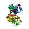

 Mass: 96.063 Da / Num. of mol.: 2 / Source method: obtained synthetically / Formula: SO4
 Mass: 18.015 Da / Num. of mol.: 253 / Source method: isolated from a natural source / Formula: H2O
 Sample preparation
 Processing