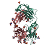
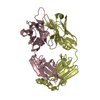
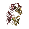
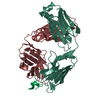
Mass: 24155.350 Da / Num. of mol.: 2 / Source method: obtained synthetically Details: RNA was prepared by in vitro transcription with T7 RNA Polymerase in Thermotoga maritima
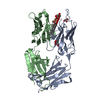
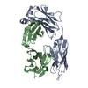
#2: Protein
polyApolymerase / A-adding enzyme
Mass: 45721.840 Da / Num. of mol.: 2 / Fragment: residues 1-390 Source method: isolated from a genetically manipulated source Source: (gene. exp.) Aquifex aeolicus (bacteria) / Plasmid: pET / Species (production host): Escherichia coli / Production host: Escherichia coli BL21(DE3) (bacteria) / Strain (production host): BL21(DE3) References: UniProt: O66728, polynucleotide adenylyltransferase
Resolution: 2.8→2.87 Å / Mean I/σ(I) obs: 4.3 / Rsym value: 0.398 / % possible all: 95.7
-
Processing
Software
Name
Classification
HKL-2000
datacollection
SCALEPACK
datascaling
MLPHARE
phasing
CNS
refinement
HKL-2000
datareduction
Refinement
Method to determine structure: MAD / Resolution: 2.8→40 Å / σ(F): 0 / Stereochemistry target values: Engh & Huber Details: This crystal has a pseudo-merohedral perfect twinning in P2, which pretends P2(1)2(1)2. beta is 90 degree since Depositors processed the data with P2(1)2(1)2, and expanded them to the P2 ...Details: This crystal has a pseudo-merohedral perfect twinning in P2, which pretends P2(1)2(1)2. beta is 90 degree since Depositors processed the data with P2(1)2(1)2, and expanded them to the P2 space group. The file contains Friedel pairs.
Rfactor
Num. reflection
Selection details
Rfree
0.2865
1945
RANDOM
Rwork
0.2297
-
-
all
-
39038
-
obs
-
39038
-
Displacement parameters
Biso mean: 87.8 Å2
Refine analyze
Free
Obs
Luzzati coordinate error
0.41 Å
0.36 Å
Luzzati d res low
-
5 Å
Luzzati sigma a
0.55 Å
0.42 Å
Refinement step
Cycle: LAST / Resolution: 2.8→40 Å
Protein
Nucleic acid
Ligand
Solvent
Total
Num. atoms
5794
1384
62
79
7319
Refine LS restraints
Refine-ID
Type
Dev ideal
X-RAY DIFFRACTION
c_bond_d
0.012
X-RAY DIFFRACTION
c_angle_deg
1.52
X-RAY DIFFRACTION
c_dihedral_angle_d
22.1
X-RAY DIFFRACTION
c_improper_angle_d
1.23
LS refinement shell
Resolution: 2.8→2.9 Å
Rfactor
Num. reflection
% reflection
Rfree
0.3263
180
-
Rwork
0.3013
-
-
obs
-
-
95 %
+
About Yorodumi
-
News
-
Feb 9, 2022. New format data for meta-information of EMDB entries
New format data for meta-information of EMDB entries
Version 3 of the EMDB header file is now the official format.
The previous official version 1.9 will be removed from the archive.
In the structure databanks used in Yorodumi, some data are registered as the other names, "COVID-19 virus" and "2019-nCoV". Here are the details of the virus and the list of structure data.
Jan 31, 2019. EMDB accession codes are about to change! (news from PDBe EMDB page)
EMDB accession codes are about to change! (news from PDBe EMDB page)
The allocation of 4 digits for EMDB accession codes will soon come to an end. Whilst these codes will remain in use, new EMDB accession codes will include an additional digit and will expand incrementally as the available range of codes is exhausted. The current 4-digit format prefixed with “EMD-” (i.e. EMD-XXXX) will advance to a 5-digit format (i.e. EMD-XXXXX), and so on. It is currently estimated that the 4-digit codes will be depleted around Spring 2019, at which point the 5-digit format will come into force.
The EM Navigator/Yorodumi systems omit the EMD- prefix.
Related info.:Q: What is EMD? / ID/Accession-code notation in Yorodumi/EM Navigator
Yorodumi is a browser for structure data from EMDB, PDB, SASBDB, etc.
This page is also the successor to EM Navigator detail page, and also detail information page/front-end page for Omokage search.
The word "yorodu" (or yorozu) is an old Japanese word meaning "ten thousand". "mi" (miru) is to see.
Related info.:EMDB / PDB / SASBDB / Comparison of 3 databanks / Yorodumi Search / Aug 31, 2016. New EM Navigator & Yorodumi / Yorodumi Papers / Jmol/JSmol / Function and homology information / Changes in new EM Navigator and Yorodumi
 Movie
Movie Controller

 Yorodumi
 Open data
 Basic information
 Components
 Keywords
 Function and homology information
 Aquifex aeolicus (bacteria)
 X-RAY DIFFRACTION /
  Authors
 Citation
 Structure visualization
 Downloads & links
 Other downloads


 PDBj
PDBj


 Assembly
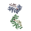
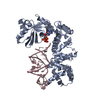
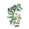

 Mass: 505.208 Da / Num. of mol.: 2 / Source method: obtained synthetically / Formula: C11H18N5O12P3 / Comment: AMP-CPP, energy-carrying molecule analogue*YM
 Mass: 18.015 Da / Num. of mol.: 79 / Source method: isolated from a natural source / Formula: H2O
 Sample preparation
 / Beamline: BL41XU / Wavelength: 1.000, 0.9793, 0.9795, 0.9843, 0.9743
 Processing