[English] 日本語
Yorodumi
- PDB-4olt: Chitosanase complex structure -

+
Open data


ID or keywords:

Loading...

-
Basic information

Entry
Database: PDB / ID: 4olt
TitleChitosanase complex structure
ComponentsChitosanase
KeywordsHYDROLASE / chitosanase / Glycoside hydrolase / chitosan
Function / homology
Function and homology information


chitosanase / chitosanase activity / carbohydrate metabolic process / extracellular region
Similarity search - Function
Chitosanase; Chain A, domain 2 / Chitosanase, subunit A, domain 2 / Chitosanases families 46 and 80 active sites signature. / Chitosanase, subunit A; domain 1 / Chitosanase, subunit A, domain 1 / Glycoside hydrolase, family 46, N-terminal / Glycosyl hydrolase family 46 / Glycoside hydrolase, family 46 / Lysozyme-like domain superfamily / Prokaryotic membrane lipoprotein lipid attachment site profile. ...Chitosanase; Chain A, domain 2 / Chitosanase, subunit A, domain 2 / Chitosanases families 46 and 80 active sites signature. / Chitosanase, subunit A; domain 1 / Chitosanase, subunit A, domain 1 / Glycoside hydrolase, family 46, N-terminal / Glycosyl hydrolase family 46 / Glycoside hydrolase, family 46 / Lysozyme-like domain superfamily / Prokaryotic membrane lipoprotein lipid attachment site profile. / Up-down Bundle / 2-Layer Sandwich / Mainly Alpha / Alpha Beta
Similarity search - Domain/homology
Biological speciesPseudomonas sp. LL2
MethodX-RAY DIFFRACTION / SYNCHROTRON / MOLECULAR REPLACEMENT / Resolution: 1.59 Å
AuthorsLiu, W.Z. / Lyu, Q.Q. / Han, B.Q.
CitationJournal: Biochem.J. / Year: 2014
Title: Structural insights into the substrate-binding mechanism for a novel chitosanase.
Authors: Lyu, Q. / Wang, S. / Xu, W. / Han, B. / Liu, W. / Jones, D.N. / Liu, W.
History
DepositionJan 25, 2014Deposition site: RCSB / Processing site: RCSB
Revision 1.0Apr 30, 2014Provider: repository / Type: Initial release
Revision 1.1Aug 6, 2014Group: Database references
Revision 2.0Jul 29, 2020Group: Advisory / Atomic model ...Advisory / Atomic model / Data collection / Database references / Derived calculations / Structure summary
Category: atom_site / atom_site_anisotrop ...atom_site / atom_site_anisotrop / chem_comp / entity / pdbx_branch_scheme / pdbx_chem_comp_identifier / pdbx_entity_branch / pdbx_entity_branch_descriptor / pdbx_entity_branch_link / pdbx_entity_branch_list / pdbx_entity_nonpoly / pdbx_nonpoly_scheme / pdbx_struct_assembly_gen / pdbx_validate_close_contact / struct_asym / struct_conn / struct_ref_seq_dif / struct_site / struct_site_gen
Item: _atom_site.B_iso_or_equiv / _atom_site.Cartn_x ..._atom_site.B_iso_or_equiv / _atom_site.Cartn_x / _atom_site.Cartn_y / _atom_site.Cartn_z / _atom_site.auth_asym_id / _atom_site.auth_atom_id / _atom_site.auth_comp_id / _atom_site.auth_seq_id / _atom_site.label_alt_id / _atom_site.label_asym_id / _atom_site.label_atom_id / _atom_site.label_comp_id / _atom_site.label_entity_id / _atom_site.occupancy / _atom_site.type_symbol / _atom_site_anisotrop.U[1][1] / _atom_site_anisotrop.U[1][2] / _atom_site_anisotrop.U[1][3] / _atom_site_anisotrop.U[2][2] / _atom_site_anisotrop.U[2][3] / _atom_site_anisotrop.U[3][3] / _atom_site_anisotrop.pdbx_auth_asym_id / _atom_site_anisotrop.pdbx_auth_atom_id / _atom_site_anisotrop.pdbx_auth_comp_id / _atom_site_anisotrop.pdbx_auth_seq_id / _atom_site_anisotrop.pdbx_label_alt_id / _atom_site_anisotrop.pdbx_label_asym_id / _atom_site_anisotrop.pdbx_label_atom_id / _atom_site_anisotrop.pdbx_label_comp_id / _atom_site_anisotrop.type_symbol / _chem_comp.name / _chem_comp.type / _entity.formula_weight / _entity.pdbx_description / _entity.pdbx_number_of_molecules / _entity.type / _pdbx_struct_assembly_gen.asym_id_list / _struct_ref_seq_dif.details
Description: Carbohydrate remediation / Provider: repository / Type: Remediation
Revision 2.1Sep 20, 2023Group: Data collection / Database references ...Data collection / Database references / Refinement description / Structure summary
Category: chem_comp / chem_comp_atom ...chem_comp / chem_comp_atom / chem_comp_bond / database_2 / pdbx_initial_refinement_model
Item: _chem_comp.pdbx_synonyms / _database_2.pdbx_DOI / _database_2.pdbx_database_accession

-
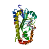
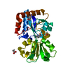
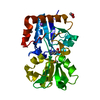
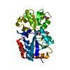
Structure visualization

Structure viewerMolecule:
MolmilJmol/JSmol

Downloads & links

-
Assembly

Deposited unit
A: Chitosanase
B: Chitosanase
hetero molecules


Theoretical massNumber of molelcules
Total (without water)55,4587
Polymers53,2112
Non-polymers2,2465
Water7,170398
1
A: Chitosanase
hetero molecules


Theoretical massNumber of molelcules
Total (without water)27,7754
Polymers26,6061
Non-polymers1,1693
Water181
TypeNameSymmetry operationNumber
identity operation1_555x,y,z1
MethodPISA
2
B: Chitosanase
hetero molecules


Theoretical massNumber of molelcules
Total (without water)27,6833
Polymers26,6061
Non-polymers1,0772
Water181
TypeNameSymmetry operationNumber
identity operation1_555x,y,z1
MethodPISA
Unit cell
Length a, b, c (Å)62.023, 40.705, 104.794
Angle α, β, γ (deg.)90.00, 106.53, 90.00
Int Tables number4
Space group name H-MP1211

-
Components

#1: Protein Chitosanase


Mass: 26605.674 Da / Num. of mol.: 2
Source method: isolated from a genetically manipulated source
Source: (gene. exp.) Pseudomonas sp. LL2(2010) (bacteria) / Gene: CHI, chitosanase OU01 / Plasmid: pGEX6p-1 / Production host: Escherichia coli (E. coli) / Strain (production host): BL21(DE3) / References: UniProt: E1AXU1, chitosanase
#2: Polysaccharide 2-amino-2-deoxy-beta-D-glucopyranose-(1-4)-2-amino-2-deoxy-beta-D-glucopyranose-(1-4)-2-amino-2- ...2-amino-2-deoxy-beta-D-glucopyranose-(1-4)-2-amino-2-deoxy-beta-D-glucopyranose-(1-4)-2-amino-2-deoxy-beta-D-glucopyranose-(1-4)-2-amino-2-deoxy-beta-D-glucopyranose-(1-4)-2-amino-2-deoxy-beta-D-glucopyranose-(1-4)-2-amino-2-deoxy-beta-D-glucopyranose


Type: oligosaccharide / Mass: 984.950 Da / Num. of mol.: 2
Source method: isolated from a genetically manipulated source
DescriptorTypeProgram
DGlcpNb1-4DGlcpNb1-4DGlcpNb1-4DGlcpNb1-4DGlcpNb1-4DGlcpNb1-ROHGlycam Condensed SequenceGMML 1.0
WURCS=2.0/1,6,5/[a2122h-1b_1-5_2*N]/1-1-1-1-1-1/a4-b1_b4-c1_c4-d1_d4-e1_e4-f1WURCSPDB2Glycan 1.1.0
[][b-D-GlcpN]{[(4+1)][b-D-GlcpN]{[(4+1)][b-D-GlcpN]{[(4+1)][b-D-GlcpN]{[(4+1)][b-D-GlcpN]{[(4+1)][b-D-GlcpN]{}}}}}}LINUCSPDB-CARE
#3: Chemical ChemComp-GOL / GLYCEROL / GLYCERIN / PROPANE-1,2,3-TRIOL


Mass: 92.094 Da / Num. of mol.: 3 / Source method: obtained synthetically / Formula: C3H8O3
#4: Water ChemComp-HOH / water


Mass: 18.015 Da / Num. of mol.: 398 / Source method: isolated from a natural source / Formula: H2O
Sequence detailsAUTHORS HAVE INDICATED THAT THE CHROMOSOMAL DNA OF MICROBACTERIUM SP. WAS ISOLATED AS THE PCR ...AUTHORS HAVE INDICATED THAT THE CHROMOSOMAL DNA OF MICROBACTERIUM SP. WAS ISOLATED AS THE PCR TEMPLATE USING BACTERIA DNA KIT (TIANGEN). BASED ON THE SUBMITTED SEQUENCE OF MICROBACTERIUM SP. OU01 CHITOSANASE GENE (GENBANK ACCESSION: EF159153), TWO PRIMERS WERE DESIGNED AS FOLLOWING: P1, 5 -TCCTCGGTTGGAGCAGCAG-3 ; P2, 5 -GTGGATTCAGCCCAGACCG-3 . COMPARED WITH CHITOSANASE (GENBANK ACCESSION: ABM91442), THE AMINO ACID SEQUENCE ENCODED BY THE CLONED CHITOSANASE OU01 GENE HAD THREE DIFFERENT RESIDUES. (COMPARED TO THE PREVIOUS SUBMITTED CHITOSANASE OU01 SEQUENCE (GENBANK ACCESSION: ABM91442), RESIDUES IN POSITION 68, 91 AND 237 ARE TYR, ASP AND TYR, RESPECTIVELY, HOWEVER, IN THIS STUDY, THE CORRESPONDING RESIDUES ARE HIS, GLY AND PHE.

-
Experimental details

-
Experiment

ExperimentMethod: X-RAY DIFFRACTION / Number of used crystals: 1

-
Sample preparation

CrystalDensity Matthews: 2.38 Å3/Da / Density % sol: 48.39 %
Crystal growTemperature: 288 K
Details: 0.05 M KH2PO4, 23% PEG 8000, VAPOR DIFFUSION, HANGING DROP, temperature 288K

-
Data collection

DiffractionMean temperature: 77 K
Diffraction sourceSource: SYNCHROTRON / Site: SSRF / Beamline: BL17U / Wavelength: 0.97869
DetectorType: ADSC QUANTUM 315r / Detector: CCD
RadiationProtocol: SINGLE WAVELENGTH / Monochromatic (M) / Laue (L): M / Scattering type: x-ray
Radiation wavelengthWavelength: 0.97869 Å / Relative weight: 1
ReflectionResolution: 1.59→33.9 Å / Num. obs: 63744 / % possible obs: 99.4 % / Redundancy: 6.5 % / Biso Wilson estimate: 15.7 Å2 / Rmerge(I) obs: 0.25 / Rsym value: 0.264 / Net I/σ(I): 12.7
Reflection shellResolution: 1.59→1.64 Å / Redundancy: 5.4 % / Mean I/σ(I) obs: 1.6 / % possible all: 99.7

-
Processing

Software
NameVersionClassification
CBASSdata collection
PHASERphasing
REFMAC5.7.0032refinement
HKL-2000data reduction
HKL-2000data scaling
RefinementMethod to determine structure: MOLECULAR REPLACEMENT
Starting model: PDB ENTRY 1CHK
Resolution: 1.59→33.9 Å / Cor.coef. Fo:Fc: 0.963 / Cor.coef. Fo:Fc free: 0.935 / SU B: 4.495 / SU ML: 0.07 / Cross valid method: THROUGHOUT / ESU R: 0.107 / ESU R Free: 0.095 / Stereochemistry target values: MAXIMUM LIKELIHOOD
RfactorNum. reflection% reflectionSelection details
Rfree0.226 3392 5.1 %RANDOM
Rwork0.161 ---
obs0.164 63744 99.4 %-
all-67136 --
Solvent computationIon probe radii: 0.8 Å / Shrinkage radii: 0.8 Å / VDW probe radii: 1.2 Å / Solvent model: MASK
Displacement parametersBiso mean: 21.54 Å2
Baniso -1Baniso -2Baniso -3
1-0.25 Å2-0 Å20 Å2
2---0.13 Å2-0 Å2
3----0.1 Å2
Refinement stepCycle: LAST / Resolution: 1.59→33.9 Å
ProteinNucleic acidLigandSolventTotal
Num. atoms3592 0 152 398 4142
Refine LS restraints
Refine-IDTypeDev idealDev ideal targetNumber
X-RAY DIFFRACTIONr_bond_refined_d0.020.0193940
X-RAY DIFFRACTIONr_bond_other_d
X-RAY DIFFRACTIONr_angle_refined_deg1.8141.9855358
X-RAY DIFFRACTIONr_angle_other_deg
X-RAY DIFFRACTIONr_dihedral_angle_1_deg5.2785495
X-RAY DIFFRACTIONr_dihedral_angle_2_deg35.87224.4200
X-RAY DIFFRACTIONr_dihedral_angle_3_deg13.37515542
X-RAY DIFFRACTIONr_dihedral_angle_4_deg20.0241528
X-RAY DIFFRACTIONr_chiral_restr0.1250.2594
X-RAY DIFFRACTIONr_gen_planes_refined0.0110.0213082
X-RAY DIFFRACTIONr_gen_planes_other
X-RAY DIFFRACTIONr_nbd_refined
X-RAY DIFFRACTIONr_nbd_other
X-RAY DIFFRACTIONr_nbtor_refined
X-RAY DIFFRACTIONr_nbtor_other
X-RAY DIFFRACTIONr_xyhbond_nbd_refined
X-RAY DIFFRACTIONr_xyhbond_nbd_other
X-RAY DIFFRACTIONr_metal_ion_refined
X-RAY DIFFRACTIONr_metal_ion_other
X-RAY DIFFRACTIONr_symmetry_vdw_refined
X-RAY DIFFRACTIONr_symmetry_vdw_other
X-RAY DIFFRACTIONr_symmetry_hbond_refined
X-RAY DIFFRACTIONr_symmetry_hbond_other
X-RAY DIFFRACTIONr_symmetry_metal_ion_refined
X-RAY DIFFRACTIONr_symmetry_metal_ion_other
X-RAY DIFFRACTIONr_mcbond_it3.511.841935
X-RAY DIFFRACTIONr_mcbond_other
X-RAY DIFFRACTIONr_mcangle_it4.0032.7722430
X-RAY DIFFRACTIONr_mcangle_other
X-RAY DIFFRACTIONr_scbond_it5.1312.1262005
X-RAY DIFFRACTIONr_scbond_other
X-RAY DIFFRACTIONr_scangle_it
X-RAY DIFFRACTIONr_scangle_other
X-RAY DIFFRACTIONr_long_range_B_refined5.3117.1456667
X-RAY DIFFRACTIONr_long_range_B_other
X-RAY DIFFRACTIONr_rigid_bond_restr5.56833940
X-RAY DIFFRACTIONr_sphericity_free31.6075145
X-RAY DIFFRACTIONr_sphericity_bonded16.64554095
LS refinement shellResolution: 1.59→1.64 Å / Total num. of bins used: 20
RfactorNum. reflection% reflection
Rfree0.366 220 -
Rwork0.24 4493 -
obs--94.91 %

+
About Yorodumi

-
News

-
Feb 9, 2022. New format data for meta-information of EMDB entries

New format data for meta-information of EMDB entries

  • Version 3 of the EMDB header file is now the official format.
  • The previous official version 1.9 will be removed from the archive.

Related info.:EMDB header

External links:wwPDB to switch to version 3 of the EMDB data model

-
Aug 12, 2020. Covid-19 info

Covid-19 info

URL: https://pdbj.org/emnavi/covid19.php

New page: Covid-19 featured information page in EM Navigator.

Related info.:Covid-19 info / Mar 5, 2020. Novel coronavirus structure data

+
Mar 5, 2020. Novel coronavirus structure data

Novel coronavirus structure data

Related info.:Yorodumi Speices / Aug 12, 2020. Covid-19 info

External links:COVID-19 featured content - PDBj / Molecule of the Month (242):Coronavirus Proteases

+
Jan 31, 2019. EMDB accession codes are about to change! (news from PDBe EMDB page)

EMDB accession codes are about to change! (news from PDBe EMDB page)

  • The allocation of 4 digits for EMDB accession codes will soon come to an end. Whilst these codes will remain in use, new EMDB accession codes will include an additional digit and will expand incrementally as the available range of codes is exhausted. The current 4-digit format prefixed with “EMD-” (i.e. EMD-XXXX) will advance to a 5-digit format (i.e. EMD-XXXXX), and so on. It is currently estimated that the 4-digit codes will be depleted around Spring 2019, at which point the 5-digit format will come into force.
  • The EM Navigator/Yorodumi systems omit the EMD- prefix.

Related info.:Q: What is EMD? / ID/Accession-code notation in Yorodumi/EM Navigator

External links:EMDB Accession Codes are Changing Soon! / Contact to PDBj

+
Jul 12, 2017. Major update of PDB

Major update of PDB

  • wwPDB released updated PDB data conforming to the new PDBx/mmCIF dictionary.
  • This is a major update changing the version number from 4 to 5, and with Remediation, in which all the entries are updated.
  • In this update, many items about electron microscopy experimental information are reorganized (e.g. em_software).
  • Now, EM Navigator and Yorodumi are based on the updated data.

External links:wwPDB Remediation / Enriched Model Files Conforming to OneDep Data Standards Now Available in the PDB FTP Archive

-
Yorodumi

Thousand views of thousand structures

  • Yorodumi is a browser for structure data from EMDB, PDB, SASBDB, etc.
  • This page is also the successor to EM Navigator detail page, and also detail information page/front-end page for Omokage search.
  • The word "yorodu" (or yorozu) is an old Japanese word meaning "ten thousand". "mi" (miru) is to see.

Related info.:EMDB / PDB / SASBDB / Comparison of 3 databanks / Yorodumi Search / Aug 31, 2016. New EM Navigator & Yorodumi / Yorodumi Papers / Jmol/JSmol / Function and homology information / Changes in new EM Navigator and Yorodumi

Read more