 Movie
Movie Controller


+ Open data

- Basic information
| Entry | Database: PDB / ID: 1chk | ||||||
|---|---|---|---|---|---|---|---|
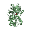
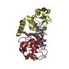
| Title | STREPTOMYCES N174 CHITOSANASE PH5.5 298K | ||||||
 Components Components | CHITOSANASE | ||||||
 Keywords Keywords | HYDROLASE (O-GLYCOSYL) / ANTI-FUNGAL PROTEIN / HYDROLASE / O-GLYCOSYL | ||||||
| Function / homology |  Function and homology information Function and homology informationchitosanase / chitosanase activity / carbohydrate metabolic process / extracellular region Similarity search - Function | ||||||
| Biological species |  Streptomyces sp. (bacteria) Streptomyces sp. (bacteria) | ||||||
| Method |  X-RAY DIFFRACTION / Resolution: 2.4 Å X-RAY DIFFRACTION / Resolution: 2.4 Å | ||||||
 Authors Authors | Marcotte, E.M. / Robertus, J.D. | ||||||
 Citation Citation | Journal: Nat.Struct.Biol. / Year: 1996 Title: X-ray structure of an anti-fungal chitosanase from streptomyces N174. Authors: Marcotte, E.M. / Monzingo, A.F. / Ernst, S.R. / Brzezinski, R. / Robertus, J.D. #1: Journal: J.Mol.Biol. / Year: 1993Title: Crystallization of a Chitosanase from Streptomyces N174 Authors: Marcotte, E. / Hart, P.J. / Boucher, I. / Brzezinski, R. / Robertus, J.D. | ||||||
| History |
|
- Structure visualization
| Structure viewer | Molecule: MolmilJmol/JSmol |
|---|
- Downloads & links
-Download
| PDBx/mmCIF format | 1chk.cif.gz | 99.9 KB | Display | PDBx/mmCIF format |
|---|---|---|---|---|
| PDB format | pdb1chk.ent.gz | 77.7 KB | Display | PDB format |
| PDBx/mmJSON format | 1chk.json.gz | Tree view | PDBx/mmJSON format | |
| Others |  Other downloads Other downloads |
-Validation report
| Arichive directory | https://data.pdbj.org/pub/pdb/validation_reports/ch/1chkftp://data.pdbj.org/pub/pdb/validation_reports/ch/1chk | HTTPS FTP |
|---|
-Related structure data
| Similar structure data |
|---|


-Links
 PDBj
PDBj- Assembly
| Deposited unit | 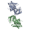
| ||||||||
|---|---|---|---|---|---|---|---|---|---|
| 1 | 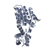
| ||||||||
| 2 | 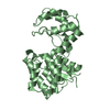
| ||||||||
| Unit cell |
| ||||||||
| Noncrystallographic symmetry (NCS) | NCS oper: (Code: given Matrix: (-0.593133, -0.777506, -0.208994), Vector: |
-Components
| #1: Protein | Mass: 25848.768 Da / Num. of mol.: 2 Source method: isolated from a genetically manipulated source Source: (gene. exp.) Streptomyces sp. (bacteria) / Strain: N174 / Description: SECRETED (MATURE PEPTIDE) / Gene: CSN / Plasmid: PRL270 / Gene (production host): CSN / Production host: Streptomyces lividans (bacteria) / References: UniProt: P33665#2: Water | ChemComp-HOH / |  Mass: 18.015 Da / Num. of mol.: 51 / Source method: isolated from a natural source / Formula: H2O Mass: 18.015 Da / Num. of mol.: 51 / Source method: isolated from a natural source / Formula: H2O |
|---|
-Experimental details
-Experiment
| Experiment | Method: X-RAY DIFFRACTION |
|---|
- Sample preparation
| Crystal | Density Matthews: 2.44 Å3/Da / Density % sol: 49.6 % | |||||||||||||||||||||||||||||||||||
|---|---|---|---|---|---|---|---|---|---|---|---|---|---|---|---|---|---|---|---|---|---|---|---|---|---|---|---|---|---|---|---|---|---|---|---|---|
| Crystal grow | pH: 5.5 / Details: pH 5.5 | |||||||||||||||||||||||||||||||||||
| Crystal | *PLUS | |||||||||||||||||||||||||||||||||||
| Crystal grow | *PLUS Method: vapor diffusion, hanging drop / Details: Marcotte, E., (1993) J.Mol.Biol., 232, 995. | |||||||||||||||||||||||||||||||||||
| Components of the solutions | *PLUS
|
-Data collection
| Diffraction | Mean temperature: 298 K |
|---|---|
| Diffraction source | Wavelength: 1.5418 |
| Detector | Type: XUONG-HAMLIN MULTIWIRE / Detector: AREA DETECTOR / Date: Sep 29, 1993 |
| Radiation | Monochromator: GRAPHITE(002) / Monochromatic (M) / Laue (L): M / Scattering type: x-ray |
| Radiation wavelength | Wavelength: 1.5418 Å / Relative weight: 1 |
| Reflection | Resolution: 2.4→50 Å / Num. obs: 21903 / % possible obs: 98 % / Observed criterion σ(I): 0 / Redundancy: 5.5 % / Rmerge(I) obs: 0.0885 |
| Reflection | *PLUS Highest resolution: 2.4 Å / % possible obs: 98.7 % / Num. measured all: 121478 |
- Processing
| Software |
| ||||||||||||||||||||||||||||||||||||||||||||||||||||||||||||
|---|---|---|---|---|---|---|---|---|---|---|---|---|---|---|---|---|---|---|---|---|---|---|---|---|---|---|---|---|---|---|---|---|---|---|---|---|---|---|---|---|---|---|---|---|---|---|---|---|---|---|---|---|---|---|---|---|---|---|---|---|---|
| Refinement | Resolution: 2.4→5 Å / σ(F): 4
| ||||||||||||||||||||||||||||||||||||||||||||||||||||||||||||
| Refinement step | Cycle: LAST / Resolution: 2.4→5 Å
| ||||||||||||||||||||||||||||||||||||||||||||||||||||||||||||
| Refine LS restraints |
| ||||||||||||||||||||||||||||||||||||||||||||||||||||||||||||
| Software | *PLUS Name: X-PLOR / Version: 3.1 / Classification: refinement | ||||||||||||||||||||||||||||||||||||||||||||||||||||||||||||
| Refinement | *PLUS % reflection Rfree: 0.236 % / Rfactor obs: 0.1809 / Rfactor Rfree: 0.1748 / Rfactor Rwork: 0.1809 | ||||||||||||||||||||||||||||||||||||||||||||||||||||||||||||
| Solvent computation | *PLUS | ||||||||||||||||||||||||||||||||||||||||||||||||||||||||||||
| Displacement parameters | *PLUS | ||||||||||||||||||||||||||||||||||||||||||||||||||||||||||||
| Refine LS restraints | *PLUS
|
