 Movie
Movie Controller

[English] 日本語
 Yorodumi
- PDB-6lln: citronellol catabolism dehydrogenase (AtuB) [Pseudomonas aerugino... -
+ Open data

- Basic information
| Entry | Database: PDB / ID: 6lln | ||||||
|---|---|---|---|---|---|---|---|
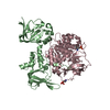
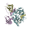
| Title | citronellol catabolism dehydrogenase (AtuB) [Pseudomonas aeruginosa PAO1] | ||||||
 Components Components | Putative dehydrogenase involved in catabolism of citronellol | ||||||
 Keywords Keywords | OXIDOREDUCTASE / Acyclic terpene utilization pathway / AtuB / Short-chain dehydrogenase/reductase(SDR) family. | ||||||
| Function / homology |  Function and homology information Function and homology information | ||||||
| Biological species |   Pseudomonas aeruginosa (bacteria) Pseudomonas aeruginosa (bacteria) | ||||||
| Method |  X-RAY DIFFRACTION / SYNCHROTRON / MOLECULAR REPLACEMENT / Resolution: 1.8 Å X-RAY DIFFRACTION / SYNCHROTRON / MOLECULAR REPLACEMENT / Resolution: 1.8 Å | ||||||
 Authors Authors | Zhang, Q. / Bartlam, M. | ||||||
| Funding support |  China, 1items China, 1items
| ||||||
 Citation Citation | Journal: Biochem.Biophys.Res.Commun. / Year: 2020 Title: Structural characterization of the Pseudomonas aeruginosa dehydrogenase AtuB involved in citronellol and geraniol catabolism. Authors: Chen, Y. / Jia, H. / Liang, Y. / Zhang, H. / Che, S. / Liu, R. / Zhang, Q. / Bartlam, M. | ||||||
| History |
|
- Structure visualization
| Structure viewer | Molecule: MolmilJmol/JSmol |
|---|
- Downloads & links
-Download
| PDBx/mmCIF format | 6lln.cif.gz | 61.2 KB | Display | PDBx/mmCIF format |
|---|---|---|---|---|
| PDB format | pdb6lln.ent.gz | 42.6 KB | Display | PDB format |
| PDBx/mmJSON format | 6lln.json.gz | Tree view | PDBx/mmJSON format | |
| Others |  Other downloads Other downloads |
-Validation report
| Arichive directory | https://data.pdbj.org/pub/pdb/validation_reports/ll/6llnftp://data.pdbj.org/pub/pdb/validation_reports/ll/6lln | HTTPS FTP |
|---|
-Related structure data
| Related structure data |  1yxmS S: Starting model for refinement |
|---|---|
| Similar structure data |


-Links
 PDBj
PDBj

- Assembly
| Deposited unit | 
| ||||||||||||
|---|---|---|---|---|---|---|---|---|---|---|---|---|---|
| 1 | 
| ||||||||||||
| Unit cell |
| ||||||||||||
| Components on special symmetry positions |
|
-Components
| #1: Protein | Mass: 30786.854 Da / Num. of mol.: 1 Source method: isolated from a genetically manipulated source Source: (gene. exp.) Pseudomonas aeruginosa (strain ATCC 15692 / DSM 22644 / CIP 104116 / JCM 14847 / LMG 12228 / 1C / PRS 101 / PAO1) (bacteria)Strain: ATCC 15692 / DSM 22644 / CIP 104116 / JCM 14847 / LMG 12228 / 1C / PRS 101 / PAO1 Gene: atuB, PA2887 / Production host: |
|---|---|
| #2: Water | ChemComp-HOH /  Mass: 18.015 Da / Num. of mol.: 129 / Source method: isolated from a natural source / Formula: H2O Mass: 18.015 Da / Num. of mol.: 129 / Source method: isolated from a natural source / Formula: H2O |
-Experimental details
-Experiment
| Experiment | Method: X-RAY DIFFRACTION / Number of used crystals: 1 |
|---|
- Sample preparation
| Crystal | Density Matthews: 2.07 Å3/Da / Density % sol: 40.7 % |
|---|---|
| Crystal grow | Temperature: 293 K / Method: vapor diffusion, sitting drop / pH: 6 Details: 0.05 M calcium chloride dihydrate, 0.1 M MES monohydrate pH 6.0, 45% PEG200 |
-Data collection
| Diffraction | Mean temperature: 100 K / Serial crystal experiment: N |
|---|---|
| Diffraction source | Source: SYNCHROTRON / Site: SSRF / Beamline: BL18U1 / Wavelength: 0.9793 Å |
| Detector | Type: DECTRIS PILATUS3 6M / Detector: PIXEL / Date: Mar 23, 2018 |
| Radiation | Protocol: SINGLE WAVELENGTH / Monochromatic (M) / Laue (L): M / Scattering type: x-ray |
| Radiation wavelength | Wavelength: 0.9793 Å / Relative weight: 1 |
| Reflection | Resolution: 1.8→50 Å / Num. obs: 23804 / % possible obs: 99.8 % / Redundancy: 12.9 % / Biso Wilson estimate: 16.44 Å2 / CC1/2: 0.99 / Rmerge(I) obs: 0.072 / Rpim(I) all: 0.021 / Net I/σ(I): 31.58 |
| Reflection shell | Resolution: 1.8→1.83 Å / Redundancy: 11.4 % / Rmerge(I) obs: 0.221 / Num. unique obs: 1155 / CC1/2: 0.99 / Rpim(I) all: 0.066 / % possible all: 98 |
- Processing
| Software |
| ||||||||||||||||||||||||
|---|---|---|---|---|---|---|---|---|---|---|---|---|---|---|---|---|---|---|---|---|---|---|---|---|---|
| Refinement | Method to determine structure: MOLECULAR REPLACEMENT Starting model: 1YXM Resolution: 1.8→49.08 Å / Cross valid method: FREE R-VALUE
| ||||||||||||||||||||||||
| Refinement step | Cycle: LAST / Resolution: 1.8→49.08 Å
| ||||||||||||||||||||||||
| Refine LS restraints |
| ||||||||||||||||||||||||
| LS refinement shell | Resolution: 1.802→1.866 Å
|
