Resolution: 2.5→20 Å / Cor.coef. Fo:Fc: 0.949 / Cor.coef. Fo:Fc free: 0.926 / SU B: 10.961 / SU ML: 0.245 / Cross valid method: THROUGHOUT / ESU R: 2.765 / ESU R Free: 0.339 / Stereochemistry target values: MAXIMUM LIKELIHOOD Details: NADPs at the binding site of enzyme with segment ID, A, B, C, E, F and G were not included in the model, due to the poor electron densities of them.
Rfactor
Num. reflection
% reflection
Selection details
Rfree
0.26273
6738
10.1 %
RANDOM
Rwork
0.22228
-
-
-
obs
0.22644
66487
100 %
-
Solvent computation
Ion probe radii: 0.8 Å / Shrinkage radii: 0.8 Å / VDW probe radii: 1.2 Å / Solvent model: BABINET MODEL WITH MASK
 Movie
Movie Controller


 Open data
 Basic information
 Components
 Keywords
 Function and homology information
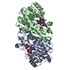
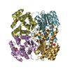
 Thermus thermophilus (bacteria)
 X-RAY DIFFRACTION /
  Authors
 Citation
 Structure visualization
 Downloads & links
 Other downloads


 PDBj
PDBj

 Assembly


 Mass: 743.405 Da / Num. of mol.: 2 / Source method: obtained synthetically / Formula: C21H28N7O17P3
 Mass: 18.015 Da / Num. of mol.: 386 / Source method: isolated from a natural source / Formula: H2O
 Sample preparation
 / Beamline: BL45XU / Wavelength: 1.02 Å
 Processing