 Movie
Movie Controller


+ Open data

- Basic information
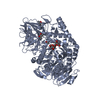
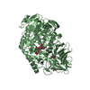
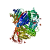
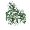
| Entry | Database: PDB / ID: 6jr6 | ||||||
|---|---|---|---|---|---|---|---|
| Title | Flavobacterium johnsoniae GH31 dextranase, FjDex31A | ||||||
 Components Components | Candidate alpha-glycosidase Glycoside hydrolase family 31 | ||||||
 Keywords Keywords | HYDROLASE / dextranase / Flavobacterium johnsoniae / dextran / isomaltose / GH31 | ||||||
| Function / homology |  Function and homology information Function and homology informationalpha-glucosidase / alpha-1,4-glucosidase activity / carbohydrate binding / carbohydrate metabolic process Similarity search - Function | ||||||
| Biological species |  Flavobacterium johnsoniae (bacteria) Flavobacterium johnsoniae (bacteria) | ||||||
| Method |  X-RAY DIFFRACTION / SYNCHROTRON / SAD / Resolution: 2 Å X-RAY DIFFRACTION / SYNCHROTRON / SAD / Resolution: 2 Å | ||||||
 Authors Authors | Tonozuka, T. | ||||||
| Funding support |  Japan, 1items Japan, 1items
| ||||||
 Citation Citation | Journal: Febs J. / Year: 2020 Title: Structural insights into polysaccharide recognition by Flavobacterium johnsoniae dextranase, a member of glycoside hydrolase family 31. Authors: Tsutsumi, K. / Gozu, Y. / Nishikawa, A. / Tonozuka, T. | ||||||
| History |
|
- Structure visualization
| Structure viewer | Molecule: MolmilJmol/JSmol |
|---|
- Downloads & links
-Download
| PDBx/mmCIF format | 6jr6.cif.gz | 706.8 KB | Display | PDBx/mmCIF format |
|---|---|---|---|---|
| PDB format | pdb6jr6.ent.gz | 568.4 KB | Display | PDB format |
| PDBx/mmJSON format | 6jr6.json.gz | Tree view | PDBx/mmJSON format | |
| Others |  Other downloads Other downloads |
-Validation report
| Arichive directory | https://data.pdbj.org/pub/pdb/validation_reports/jr/6jr6ftp://data.pdbj.org/pub/pdb/validation_reports/jr/6jr6 | HTTPS FTP |
|---|
-Related structure data


-Links
 PDBj
PDBj


- Assembly
| Deposited unit | 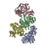
| ||||||||
|---|---|---|---|---|---|---|---|---|---|
| 1 | 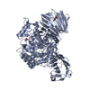
| ||||||||
| 2 | 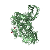
| ||||||||
| 3 | 
| ||||||||
| 4 | 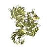
| ||||||||
| Unit cell |
|
-Components
| #1: Protein | Mass: 96182.477 Da / Num. of mol.: 4 Source method: isolated from a genetically manipulated source Source: (gene. exp.) Flavobacterium johnsoniae (strain ATCC 17061 / DSM 2064 / UW101) (bacteria)Strain: ATCC 17061 / DSM 2064 / UW101 / Gene: Fjoh_4430 / Production host: #2: Chemical | ChemComp-ACT /   Mass: 59.044 Da / Num. of mol.: 4 / Source method: obtained synthetically / Formula: C2H3O2 Mass: 59.044 Da / Num. of mol.: 4 / Source method: obtained synthetically / Formula: C2H3O2#3: Chemical | ChemComp-MPD / (   Mass: 118.174 Da / Num. of mol.: 8 / Source method: obtained synthetically / Formula: C6H14O2 / Comment: precipitant*YM Mass: 118.174 Da / Num. of mol.: 8 / Source method: obtained synthetically / Formula: C6H14O2 / Comment: precipitant*YM#4: Water | ChemComp-HOH / |  Mass: 18.015 Da / Num. of mol.: 2381 / Source method: isolated from a natural source / Formula: H2O Mass: 18.015 Da / Num. of mol.: 2381 / Source method: isolated from a natural source / Formula: H2OHas protein modification | Y | |
|---|
-Experimental details
-Experiment
| Experiment | Method: X-RAY DIFFRACTION / Number of used crystals: 1 |
|---|
- Sample preparation
| Crystal | Density Matthews: 2.92 Å3/Da / Density % sol: 57.81 % |
|---|---|
| Crystal grow | Temperature: 293 K / Method: vapor diffusion, hanging drop / pH: 8 Details: 100 mM sodium acetate buffer, 8% (w/v) polyethylene glycol 20000, and 8% 2-methyl-2,4-pentanediol |
-Data collection
| Diffraction | Mean temperature: 100 K / Serial crystal experiment: N |
|---|---|
| Diffraction source | Source: SYNCHROTRON / Site: Photon Factory / Beamline: BL-5A / Wavelength: 1 Å |
| Detector | Type: ADSC QUANTUM 315r / Detector: CCD / Date: Nov 23, 2016 |
| Radiation | Protocol: SINGLE WAVELENGTH / Monochromatic (M) / Laue (L): M / Scattering type: x-ray |
| Radiation wavelength | Wavelength: 1 Å / Relative weight: 1 |
| Reflection | Resolution: 2→48.5 Å / Num. obs: 275382 / % possible obs: 97.1 % / Redundancy: 1.8 % / Rmerge(I) obs: 0.091 / Net I/σ(I): 7.8 |
| Reflection shell | Resolution: 2→2.03 Å / Redundancy: 1.7 % / Rmerge(I) obs: 0.334 / Mean I/σ(I) obs: 1.8 / Num. unique obs: 13574 / % possible all: 96.1 |
- Processing
| Software |
| ||||||||||||||||||||||||||||||||||||||||||||||||||||||||||||
|---|---|---|---|---|---|---|---|---|---|---|---|---|---|---|---|---|---|---|---|---|---|---|---|---|---|---|---|---|---|---|---|---|---|---|---|---|---|---|---|---|---|---|---|---|---|---|---|---|---|---|---|---|---|---|---|---|---|---|---|---|---|
| Refinement | Method to determine structure: SAD / Resolution: 2→48.45 Å / Cor.coef. Fo:Fc: 0.962 / Cor.coef. Fo:Fc free: 0.945 / Cross valid method: THROUGHOUT / σ(F): 0 / ESU R: 0.164 / ESU R Free: 0.144 / Stereochemistry target values: MAXIMUM LIKELIHOOD Details: Leu308-Gln309 adopts a cis conformation yet the electron density is clearly seen HYDROGENS HAVE BEEN ADDED IN THE RIDING POSITIONS U VALUES : REFINED INDIVIDUALLY
| ||||||||||||||||||||||||||||||||||||||||||||||||||||||||||||
| Solvent computation | Ion probe radii: 0.8 Å / Shrinkage radii: 0.8 Å / VDW probe radii: 1.2 Å / Solvent model: MASK | ||||||||||||||||||||||||||||||||||||||||||||||||||||||||||||
| Displacement parameters | Biso max: 100.4 Å2 / Biso mean: 28.199 Å2 / Biso min: 14.55 Å2
| ||||||||||||||||||||||||||||||||||||||||||||||||||||||||||||
| Refinement step | Cycle: LAST / Resolution: 2→48.45 Å
| ||||||||||||||||||||||||||||||||||||||||||||||||||||||||||||
| Refine LS restraints |
| ||||||||||||||||||||||||||||||||||||||||||||||||||||||||||||
| LS refinement shell | Resolution: 2.001→2.053 Å / Rfactor Rfree error: 0 / Total num. of bins used: 20
|
