 Movie
Movie Controller


+ Open data

- Basic information
| Entry | Database: PDB / ID: 6lbf | ||||||
|---|---|---|---|---|---|---|---|
| Title | Crystal structure of FEM1B | ||||||
 Components Components | Protein fem-1 homolog B | ||||||
 Keywords Keywords | PROTEIN BINDING / ubiquitination / E3 ligase | ||||||
| Function / homology |  Function and homology information Function and homology informationregulation of ubiquitin-protein transferase activity / epithelial cell maturation involved in prostate gland development / branching involved in prostate gland morphogenesis / regulation of DNA damage checkpoint / death receptor binding / regulation of extrinsic apoptotic signaling pathway via death domain receptors / ubiquitin-dependent protein catabolic process via the C-end degron rule pathway / Cul2-RING ubiquitin ligase complex / ubiquitin ligase complex / ubiquitin-like ligase-substrate adaptor activity ...regulation of ubiquitin-protein transferase activity / epithelial cell maturation involved in prostate gland development / branching involved in prostate gland morphogenesis / regulation of DNA damage checkpoint / death receptor binding / regulation of extrinsic apoptotic signaling pathway via death domain receptors / ubiquitin-dependent protein catabolic process via the C-end degron rule pathway / Cul2-RING ubiquitin ligase complex / ubiquitin ligase complex / ubiquitin-like ligase-substrate adaptor activity / Neddylation / proteasome-mediated ubiquitin-dependent protein catabolic process / protein ubiquitination / apoptotic process / mitochondrion / nucleoplasm / metal ion binding / nucleus / cytoplasm / cytosol Similarity search - Function | ||||||
| Biological species |  Homo sapiens (human) Homo sapiens (human) | ||||||
| Method |  X-RAY DIFFRACTION / SYNCHROTRON / MAD / Resolution: 3.252 Å X-RAY DIFFRACTION / SYNCHROTRON / MAD / Resolution: 3.252 Å | ||||||
 Authors Authors | Chen, X. / Liao, S. / Xu, C. | ||||||
| Funding support |  China, 1items China, 1items
| ||||||
 Citation Citation | Journal: Nat.Chem.Biol. / Year: 2021 Title: Molecular basis for arginine C-terminal degron recognition by Cul2 FEM1 E3 ligase. Authors: Chen, X. / Liao, S. / Makaros, Y. / Guo, Q. / Zhu, Z. / Krizelman, R. / Dahan, K. / Tu, X. / Yao, X. / Koren, I. / Xu, C. | ||||||
| History |
|
- Structure visualization
| Structure viewer | Molecule: MolmilJmol/JSmol |
|---|
- Downloads & links
-Download
| PDBx/mmCIF format | 6lbf.cif.gz | 135.6 KB | Display | PDBx/mmCIF format |
|---|---|---|---|---|
| PDB format | pdb6lbf.ent.gz | 105.6 KB | Display | PDB format |
| PDBx/mmJSON format | 6lbf.json.gz | Tree view | PDBx/mmJSON format | |
| Others |  Other downloads Other downloads |
-Validation report
| Arichive directory | https://data.pdbj.org/pub/pdb/validation_reports/lb/6lbfftp://data.pdbj.org/pub/pdb/validation_reports/lb/6lbf | HTTPS FTP |
|---|
-Related structure data
| Related structure data | 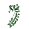 6lbgC 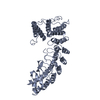 6lbnC 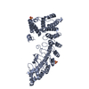 6ldpC  6le6C  6lenC  6leyC  6lf0C 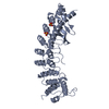 7cngC C: citing same article ( |
|---|---|
| Similar structure data |


-Links
 PDBj
PDBj

- Assembly
| Deposited unit | 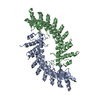
| ||||||||
|---|---|---|---|---|---|---|---|---|---|
| 1 | 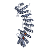
| ||||||||
| 2 | 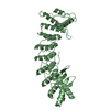
| ||||||||
| Unit cell |
|
-Components
| #1: Protein | Mass: 39459.887 Da / Num. of mol.: 2 Source method: isolated from a genetically manipulated source Source: (gene. exp.) Homo sapiens (human) / Gene: FEM1B, F1AA, KIAA0396 / Production host:  #2: Chemical |   Mass: 96.063 Da / Num. of mol.: 2 / Source method: obtained synthetically / Formula: SO4 Mass: 96.063 Da / Num. of mol.: 2 / Source method: obtained synthetically / Formula: SO4Has ligand of interest | N | |
|---|
-Experimental details
-Experiment
| Experiment | Method: X-RAY DIFFRACTION / Number of used crystals: 1 |
|---|
- Sample preparation
| Crystal | Density Matthews: 3.7 Å3/Da / Density % sol: 66.76 % |
|---|---|
| Crystal grow | Temperature: 291 K / Method: vapor diffusion, sitting drop Details: 0.1M Tris-HCl, pH 8.5, 0.1M Guanidine hydrochloride, 0.8M Potassium sodium tartrate tetrahydrate, 0.5% w/v Polyethylene glycol monomethyl ether 5000 |
-Data collection
| Diffraction | Mean temperature: 100 K / Serial crystal experiment: N |
|---|---|
| Diffraction source | Source: SYNCHROTRON / Site: SSRF / Beamline: BL17U1 / Wavelength: 0.979 Å |
| Detector | Type: ADSC QUANTUM 315r / Detector: CCD / Date: Oct 21, 2018 |
| Radiation | Protocol: MAD / Monochromatic (M) / Laue (L): M / Scattering type: x-ray |
| Radiation wavelength | Wavelength: 0.979 Å / Relative weight: 1 |
| Reflection | Resolution: 3.25→50 Å / Num. obs: 19211 / % possible obs: 100 % / Redundancy: 25.2 % / CC1/2: 1 / Rmerge(I) obs: 1.98 / Net I/σ(I): 12.3 |
| Reflection shell | Resolution: 3.25→3.37 Å / Num. unique obs: 1893 / CC1/2: 0.606 |
- Processing
| Software |
| ||||||||||||||||||||||||||||||||||||||||||||||||||||||||||||||||||||||||||||||||||||||||||
|---|---|---|---|---|---|---|---|---|---|---|---|---|---|---|---|---|---|---|---|---|---|---|---|---|---|---|---|---|---|---|---|---|---|---|---|---|---|---|---|---|---|---|---|---|---|---|---|---|---|---|---|---|---|---|---|---|---|---|---|---|---|---|---|---|---|---|---|---|---|---|---|---|---|---|---|---|---|---|---|---|---|---|---|---|---|---|---|---|---|---|---|
| Refinement | Method to determine structure: MAD / Resolution: 3.252→47.678 Å / SU ML: 0.45 / Cross valid method: THROUGHOUT / σ(F): 1.35 / Phase error: 26.98 / Stereochemistry target values: ML
| ||||||||||||||||||||||||||||||||||||||||||||||||||||||||||||||||||||||||||||||||||||||||||
| Solvent computation | Shrinkage radii: 0.9 Å / VDW probe radii: 1.11 Å / Solvent model: FLAT BULK SOLVENT MODEL | ||||||||||||||||||||||||||||||||||||||||||||||||||||||||||||||||||||||||||||||||||||||||||
| Displacement parameters | Biso max: 140 Å2 / Biso mean: 94.0095 Å2 / Biso min: 30 Å2 | ||||||||||||||||||||||||||||||||||||||||||||||||||||||||||||||||||||||||||||||||||||||||||
| Refinement step | Cycle: final / Resolution: 3.252→47.678 Å
| ||||||||||||||||||||||||||||||||||||||||||||||||||||||||||||||||||||||||||||||||||||||||||
| LS refinement shell | Refine-ID: X-RAY DIFFRACTION / Rfactor Rfree error: 0
|
