 Movie
Movie Controller

[English] 日本語
 Yorodumi
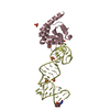
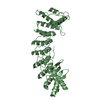
- PDB-3rmr: Crystal structure of Hyaloperonospora arabidopsidis ATR1 effector... -
+ Open data

- Basic information
| Entry | Database: PDB / ID: 3rmr | ||||||
|---|---|---|---|---|---|---|---|
| Title | Crystal structure of Hyaloperonospora arabidopsidis ATR1 effector domain | ||||||
 Components Components | Avirulence protein | ||||||
 Keywords Keywords | PROTEIN BINDING / effector / RPP1-recognized / alpha-helical / W-motif / seahorse / virulence / RPP1 / R-protein | ||||||
| Function / homology |  Function and homology information Function and homology informationeffector-mediated perturbation of host process by symbiont / host cell cytoplasm / host cell nucleus / extracellular region Similarity search - Function | ||||||
| Biological species |  Hyaloperonospora parasitica (eukaryote) Hyaloperonospora parasitica (eukaryote) | ||||||
| Method |  X-RAY DIFFRACTION / SYNCHROTRON / MAD / Resolution: 2.3 Å X-RAY DIFFRACTION / SYNCHROTRON / MAD / Resolution: 2.3 Å | ||||||
 Authors Authors | Chou, S. / Krasileva, K.V. / Holton, J.M. / Staskawicz, B.J. / Alber, T. | ||||||
 Citation Citation | Journal: To be Published Title: The Hyaloperonospora arabidopsidis ATR1 effector has distributed recognition surfaces and a structural subdomain conserved across oomycete species Authors: Chou, S. / Krasileva, K.V. / Holton, J.M. / Steinbrenner, A. / Alber, T. / Staskawicz, B.J. | ||||||
| History |
|
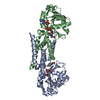
- Structure visualization
| Structure viewer | Molecule: MolmilJmol/JSmol |
|---|
- Downloads & links
-Download
| PDBx/mmCIF format | 3rmr.cif.gz | 156.9 KB | Display | PDBx/mmCIF format |
|---|---|---|---|---|
| PDB format | pdb3rmr.ent.gz | 124.2 KB | Display | PDB format |
| PDBx/mmJSON format | 3rmr.json.gz | Tree view | PDBx/mmJSON format | |
| Others |  Other downloads Other downloads |
-Validation report
| Arichive directory | https://data.pdbj.org/pub/pdb/validation_reports/rm/3rmrftp://data.pdbj.org/pub/pdb/validation_reports/rm/3rmr | HTTPS FTP |
|---|
-Related structure data
| Similar structure data |
|---|


-Links
 PDBj
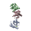
PDBj- Assembly
| Deposited unit | 
| ||||||||||||
|---|---|---|---|---|---|---|---|---|---|---|---|---|---|
| 1 | 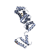
| ||||||||||||
| 2 | 
| ||||||||||||
| 3 | 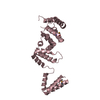
| ||||||||||||
| 4 | 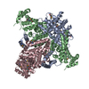
| ||||||||||||
| 5 | 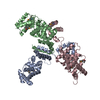
| ||||||||||||
| Unit cell |
| ||||||||||||
| Components on special symmetry positions |
|
-Components
| #1: Protein | Mass: 29676.621 Da / Num. of mol.: 3 Source method: isolated from a genetically manipulated source Source: (gene. exp.) Hyaloperonospora parasitica (eukaryote)Gene: Atr1, ATR1NdWsB / Production host:  #2: Water | ChemComp-HOH / |  Mass: 18.015 Da / Num. of mol.: 365 / Source method: isolated from a natural source / Formula: H2O Mass: 18.015 Da / Num. of mol.: 365 / Source method: isolated from a natural source / Formula: H2OHas protein modification | Y | |
|---|
-Experimental details
-Experiment
| Experiment | Method: X-RAY DIFFRACTION / Number of used crystals: 2 |
|---|
- Sample preparation
| Crystal | Density Matthews: 3.61 Å3/Da / Density % sol: 65.97 % |
|---|---|
| Crystal grow | Temperature: 291 K / Method: vapor diffusion, hanging drop / pH: 6.5 Details: Crystal growth in 1.6 M magnesium sulfate, 0.1 M MES, pH 6.5. 1:20000 seeding in 1.2 M magnesium sulfate, 0.01% acetonitrile, 0.1 M MES, pH 5.0. Dehydration in 1.5 M magnesium sulfate, 0.1 M ...Details: Crystal growth in 1.6 M magnesium sulfate, 0.1 M MES, pH 6.5. 1:20000 seeding in 1.2 M magnesium sulfate, 0.01% acetonitrile, 0.1 M MES, pH 5.0. Dehydration in 1.5 M magnesium sulfate, 0.1 M MES, pH 5.0 for 2 hours, VAPOR DIFFUSION, HANGING DROP, temperature 291K |
-Data collection
| Diffraction | Mean temperature: 100 K | ||||||||||||
|---|---|---|---|---|---|---|---|---|---|---|---|---|---|
| Diffraction source | Source: SYNCHROTRON / Site: ALS  / Beamline: 8.3.1 / Wavelength: 1.0722, 1.0631, 1.11587 / Beamline: 8.3.1 / Wavelength: 1.0722, 1.0631, 1.11587 | ||||||||||||
| Detector | Type: ADSC QUANTUM 315r / Detector: CCD / Date: May 18, 2010 | ||||||||||||
| Radiation | Monochromator: KHOZU double flat crystal / Protocol: MAD / Monochromatic (M) / Laue (L): M / Scattering type: x-ray | ||||||||||||
| Radiation wavelength |
| ||||||||||||
| Reflection | Resolution: 2.268→103.431 Å / Num. all: 60675 / Num. obs: 57939 / % possible obs: 99.4 % / Observed criterion σ(F): -3 / Observed criterion σ(I): 0 / Redundancy: 15.5 % / Rmerge(I) obs: 0.083 / Rsym value: 0.083 / Net I/σ(I): 35.55 | ||||||||||||
| Reflection shell | Resolution: 2.42→2.51 Å / Redundancy: 15.6 % / Rmerge(I) obs: 0.729 / Mean I/σ(I) obs: 4.375 / Rsym value: 0.729 / % possible all: 97.6 |
- Processing
| Software |
| |||||||||||||||||||||||||||||||||||||||||||||||||||||||||||||||||||||||||||||||||||||||||||||||||||||||||||||||||||||||||||||||||||||||||||||||||||
|---|---|---|---|---|---|---|---|---|---|---|---|---|---|---|---|---|---|---|---|---|---|---|---|---|---|---|---|---|---|---|---|---|---|---|---|---|---|---|---|---|---|---|---|---|---|---|---|---|---|---|---|---|---|---|---|---|---|---|---|---|---|---|---|---|---|---|---|---|---|---|---|---|---|---|---|---|---|---|---|---|---|---|---|---|---|---|---|---|---|---|---|---|---|---|---|---|---|---|---|---|---|---|---|---|---|---|---|---|---|---|---|---|---|---|---|---|---|---|---|---|---|---|---|---|---|---|---|---|---|---|---|---|---|---|---|---|---|---|---|---|---|---|---|---|---|---|---|---|
| Refinement | Method to determine structure: MAD / Resolution: 2.3→62.349 Å / SU ML: 0.36 / σ(F): 0 / Phase error: 26.31 / Stereochemistry target values: MLHL
| |||||||||||||||||||||||||||||||||||||||||||||||||||||||||||||||||||||||||||||||||||||||||||||||||||||||||||||||||||||||||||||||||||||||||||||||||||
| Solvent computation | Shrinkage radii: 0.83 Å / VDW probe radii: 1.1 Å / Solvent model: FLAT BULK SOLVENT MODEL / Bsol: 47.454 Å2 / ksol: 0.357 e/Å3 | |||||||||||||||||||||||||||||||||||||||||||||||||||||||||||||||||||||||||||||||||||||||||||||||||||||||||||||||||||||||||||||||||||||||||||||||||||
| Displacement parameters |
| |||||||||||||||||||||||||||||||||||||||||||||||||||||||||||||||||||||||||||||||||||||||||||||||||||||||||||||||||||||||||||||||||||||||||||||||||||
| Refinement step | Cycle: LAST / Resolution: 2.3→62.349 Å
| |||||||||||||||||||||||||||||||||||||||||||||||||||||||||||||||||||||||||||||||||||||||||||||||||||||||||||||||||||||||||||||||||||||||||||||||||||
| Refine LS restraints |
| |||||||||||||||||||||||||||||||||||||||||||||||||||||||||||||||||||||||||||||||||||||||||||||||||||||||||||||||||||||||||||||||||||||||||||||||||||
| LS refinement shell |
|
