 Movie
Movie Controller


+ Open data

- Basic information
| Entry | Database: PDB / ID: 6l1c | ||||||
|---|---|---|---|---|---|---|---|
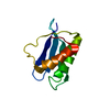
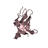
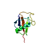
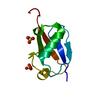
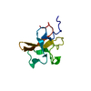
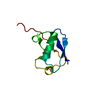
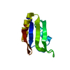
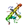
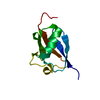
| Title | Crystal Structure Of of PHF20L1 Tudor1 Y24L mutant | ||||||
 Components Components | PHD finger protein 20-like protein 1 | ||||||
 Keywords Keywords | METAL BINDING PROTEIN / PHF20L1 / Tudor / Y24L | ||||||
| Function / homology |  Function and homology information Function and homology informationmethylation-dependent protein binding / NSL complex / Formation of WDR5-containing histone-modifying complexes / negative regulation of proteasomal ubiquitin-dependent protein catabolic process / negative regulation of protein catabolic process / regulation of transcription by RNA polymerase II / zinc ion binding / nucleoplasm Similarity search - Function | ||||||
| Biological species |  Homo sapiens (human) Homo sapiens (human) | ||||||
| Method |  X-RAY DIFFRACTION / SYNCHROTRON / MOLECULAR REPLACEMENT / Resolution: 1.58 Å X-RAY DIFFRACTION / SYNCHROTRON / MOLECULAR REPLACEMENT / Resolution: 1.58 Å | ||||||
 Authors Authors | Lv, M.Q. / Gao, J. | ||||||
 Citation Citation | Journal: J Phys Chem Lett / Year: 2020 Title: Conformational Selection in Ligand Recognition by the First Tudor Domain of PHF20L1. Authors: Lv, M. / Gao, J. / Li, M. / Ma, R. / Li, F. / Liu, Y. / Liu, M. / Zhang, J. / Yao, X. / Wu, J. / Shi, Y. / Tang, Y. / Pan, Y. / Zhang, Z. / Ruan, K. | ||||||
| History |
|
- Structure visualization
| Structure viewer | Molecule: MolmilJmol/JSmol |
|---|
- Downloads & links
-Download
| PDBx/mmCIF format | 6l1c.cif.gz | 30.2 KB | Display | PDBx/mmCIF format |
|---|---|---|---|---|
| PDB format | pdb6l1c.ent.gz | 18 KB | Display | PDB format |
| PDBx/mmJSON format | 6l1c.json.gz | Tree view | PDBx/mmJSON format | |
| Others |  Other downloads Other downloads |
-Validation report
| Arichive directory | https://data.pdbj.org/pub/pdb/validation_reports/l1/6l1cftp://data.pdbj.org/pub/pdb/validation_reports/l1/6l1c | HTTPS FTP |
|---|
-Related structure data
| Related structure data | 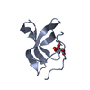 6l0xC 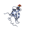 6l10C 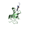 6l1fC 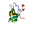 6l1iC 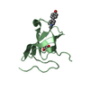 6l1pC  3sd4S S: Starting model for refinement C: citing same article ( |
|---|---|
| Similar structure data |

-Links
 PDBj
PDBj- Assembly
| Deposited unit | 
| ||||||||
|---|---|---|---|---|---|---|---|---|---|
| 1 |
| ||||||||
| Unit cell |
|
-Components
| #1: Protein | Mass: 8643.789 Da / Num. of mol.: 1 / Mutation: Y24L Source method: isolated from a genetically manipulated source Source: (gene. exp.) Homo sapiens (human) / Gene: PHF20L1, CGI-72 / Production host:  | ||||||
|---|---|---|---|---|---|---|---|
| #2: Chemical |   Mass: 96.063 Da / Num. of mol.: 3 / Source method: obtained synthetically / Formula: SO4 Mass: 96.063 Da / Num. of mol.: 3 / Source method: obtained synthetically / Formula: SO4#3: Chemical | ChemComp-GOL / |   Mass: 92.094 Da / Num. of mol.: 1 / Source method: obtained synthetically / Formula: C3H8O3 Mass: 92.094 Da / Num. of mol.: 1 / Source method: obtained synthetically / Formula: C3H8O3#4: Water | ChemComp-HOH / |  Mass: 18.015 Da / Num. of mol.: 35 / Source method: isolated from a natural source / Formula: H2O Mass: 18.015 Da / Num. of mol.: 35 / Source method: isolated from a natural source / Formula: H2OHas ligand of interest | N | |
-Experimental details
-Experiment
| Experiment | Method: X-RAY DIFFRACTION / Number of used crystals: 1 |
|---|
- Sample preparation
| Crystal | Density Matthews: 3.09 Å3/Da / Density % sol: 60.18 % |
|---|---|
| Crystal grow | Temperature: 293 K / Method: vapor diffusion, sitting drop / Details: 1.6M lithium sulfate, 0.1M Tris, PH 8.0 |
-Data collection
| Diffraction | Mean temperature: 100 K / Serial crystal experiment: N |
|---|---|
| Diffraction source | Source: SYNCHROTRON / Site: SSRF  / Beamline: BL19U1 / Wavelength: 0.979 Å / Beamline: BL19U1 / Wavelength: 0.979 Å |
| Detector | Type: ADSC QUANTUM 315r / Detector: CCD / Date: Mar 19, 2019 |
| Radiation | Protocol: SINGLE WAVELENGTH / Monochromatic (M) / Laue (L): M / Scattering type: x-ray |
| Radiation wavelength | Wavelength: 0.979 Å / Relative weight: 1 |
| Reflection twin | Operator: -k,-h,-l / Fraction: 0.488 |
| Reflection | Resolution: 1.58→37.173 Å / Num. obs: 13228 / % possible obs: 99.93 % / Redundancy: 13.2 % / Biso Wilson estimate: 16.48 Å2 / Rmerge(I) obs: 0.097 / Net I/σ(I): 37.4 |
| Reflection shell | Resolution: 1.582→1.623 Å / Rmerge(I) obs: 0.341 / Mean I/σ(I) obs: 6.3 / Num. unique obs: 956 |
- Processing
| Software |
| ||||||||||||||||||||||||||||||||||||||||||||||||||||||||||||
|---|---|---|---|---|---|---|---|---|---|---|---|---|---|---|---|---|---|---|---|---|---|---|---|---|---|---|---|---|---|---|---|---|---|---|---|---|---|---|---|---|---|---|---|---|---|---|---|---|---|---|---|---|---|---|---|---|---|---|---|---|---|
| Refinement | Method to determine structure: MOLECULAR REPLACEMENT Starting model: 3SD4 Resolution: 1.58→37.17 Å / Cor.coef. Fo:Fc: 0.953 / Cor.coef. Fo:Fc free: 0.912 / WRfactor Rfree: 0.2302 / WRfactor Rwork: 0.1911 / FOM work R set: 0.9092 / SU B: 0.858 / SU ML: 0.032 / SU R Cruickshank DPI: 0.0151 / SU Rfree: 0.0145 / Cross valid method: THROUGHOUT / σ(F): 0 / ESU R: 0.015 / ESU R Free: 0.015 / Stereochemistry target values: MAXIMUM LIKELIHOOD Details: HYDROGENS HAVE BEEN ADDED IN THE RIDING POSITIONS U VALUES : REFINED INDIVIDUALLY
| ||||||||||||||||||||||||||||||||||||||||||||||||||||||||||||
| Solvent computation | Ion probe radii: 0.8 Å / Shrinkage radii: 0.8 Å / VDW probe radii: 1.2 Å / Solvent model: MASK | ||||||||||||||||||||||||||||||||||||||||||||||||||||||||||||
| Displacement parameters | Biso max: 72.94 Å2 / Biso mean: 16.575 Å2 / Biso min: 10.03 Å2
| ||||||||||||||||||||||||||||||||||||||||||||||||||||||||||||
| Refinement step | Cycle: final / Resolution: 1.58→37.17 Å
| ||||||||||||||||||||||||||||||||||||||||||||||||||||||||||||
| Refine LS restraints |
| ||||||||||||||||||||||||||||||||||||||||||||||||||||||||||||
| LS refinement shell | Resolution: 1.582→1.623 Å / Rfactor Rfree error: 0 / Total num. of bins used: 20
|
