 Movie
Movie Controller

[English] 日本語
 Yorodumi
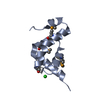
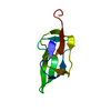
- PDB-6l1f: Crystal structure of PHF20L1 Tudor1 in complex with K142me1 DNMT1 -
+ Open data

- Basic information
| Entry | Database: PDB / ID: 6l1f | ||||||
|---|---|---|---|---|---|---|---|
| Title | Crystal structure of PHF20L1 Tudor1 in complex with K142me1 DNMT1 | ||||||
 Components Components |
| ||||||
 Keywords Keywords | METAL BINDING PROTEIN/PEPTIDE / PHF20L1 / Tudor / apo / METAL BINDING PROTEIN-PEPTIDE complex | ||||||
| Function / homology |  Function and homology information Function and homology informationmethylation-dependent protein binding / histone H3K23ub reader activity / histone H3K18ub reader activity / : / histone H3K14ub reader activity / chromosomal DNA methylation maintenance following DNA replication / negative regulation of vascular associated smooth muscle cell apoptotic process / DNA-methyltransferase activity / DNA (cytosine-5-)-methyltransferase / DNA (cytosine-5-)-methyltransferase activity ...methylation-dependent protein binding / histone H3K23ub reader activity / histone H3K18ub reader activity / : / histone H3K14ub reader activity / chromosomal DNA methylation maintenance following DNA replication / negative regulation of vascular associated smooth muscle cell apoptotic process / DNA-methyltransferase activity / DNA (cytosine-5-)-methyltransferase / DNA (cytosine-5-)-methyltransferase activity / SUMOylation of DNA methylation proteins / STAT3 nuclear events downstream of ALK signaling / NSL complex / methyl-CpG binding / negative regulation of gene expression via chromosomal CpG island methylation / DNA methylation-dependent constitutive heterochromatin formation / Formation of WDR5-containing histone-modifying complexes / lncRNA binding / pericentric heterochromatin / positive regulation of vascular associated smooth muscle cell proliferation / heterochromatin / Nuclear events stimulated by ALK signaling in cancer / negative regulation of proteasomal ubiquitin-dependent protein catabolic process / DNA methylation / replication fork / PRC2 methylates histones and DNA / Defective pyroptosis / promoter-specific chromatin binding / negative regulation of protein catabolic process / NoRC negatively regulates rRNA expression / methylation / negative regulation of gene expression / positive regulation of gene expression / regulation of transcription by RNA polymerase II / negative regulation of transcription by RNA polymerase II / DNA-templated transcription / mitochondrion / DNA binding / zinc ion binding / nucleoplasm / nucleus Similarity search - Function | ||||||
| Biological species |  Homo sapiens (human) Homo sapiens (human) | ||||||
| Method |  X-RAY DIFFRACTION / SYNCHROTRON / MOLECULAR REPLACEMENT / Resolution: 1.9 Å X-RAY DIFFRACTION / SYNCHROTRON / MOLECULAR REPLACEMENT / Resolution: 1.9 Å | ||||||
 Authors Authors | Lv, M.Q. / Gao, J. | ||||||
 Citation Citation | Journal: J Phys Chem Lett / Year: 2020 Title: Conformational Selection in Ligand Recognition by the First Tudor Domain of PHF20L1. Authors: Lv, M. / Gao, J. / Li, M. / Ma, R. / Li, F. / Liu, Y. / Liu, M. / Zhang, J. / Yao, X. / Wu, J. / Shi, Y. / Tang, Y. / Pan, Y. / Zhang, Z. / Ruan, K. | ||||||
| History |
|
- Structure visualization
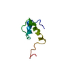
| Structure viewer | Molecule: MolmilJmol/JSmol |
|---|
- Downloads & links
-Download
| PDBx/mmCIF format | 6l1f.cif.gz | 47.5 KB | Display | PDBx/mmCIF format |
|---|---|---|---|---|
| PDB format | pdb6l1f.ent.gz | 31.2 KB | Display | PDB format |
| PDBx/mmJSON format | 6l1f.json.gz | Tree view | PDBx/mmJSON format | |
| Others |  Other downloads Other downloads |
-Validation report
| Arichive directory | https://data.pdbj.org/pub/pdb/validation_reports/l1/6l1fftp://data.pdbj.org/pub/pdb/validation_reports/l1/6l1f | HTTPS FTP |
|---|
-Related structure data
| Related structure data | 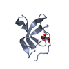 6l0xC 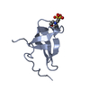 6l10C 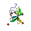 6l1cC 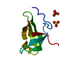 6l1iC 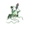 6l1pC  3sd4S S: Starting model for refinement C: citing same article ( |
|---|---|
| Similar structure data |


-Links
 PDBj
PDBj


- Assembly
| Deposited unit | 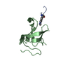
| ||||||||
|---|---|---|---|---|---|---|---|---|---|
| 1 |
| ||||||||
| Unit cell |
|
-Components
| #1: Protein/peptide | Mass: 663.703 Da / Num. of mol.: 1 / Source method: obtained synthetically / Details: MLZ represent mono-methylated lysine / Source: (synth.) Homo sapiens (human) / References: UniProt: P26358 |
|---|---|
| #2: Protein | Mass: 8693.805 Da / Num. of mol.: 1 Source method: isolated from a genetically manipulated source Source: (gene. exp.) Homo sapiens (human) / Gene: PHF20L1, CGI-72 / Production host:  |
| #3: Water | ChemComp-HOH /  Mass: 18.015 Da / Num. of mol.: 32 / Source method: isolated from a natural source / Formula: H2O Mass: 18.015 Da / Num. of mol.: 32 / Source method: isolated from a natural source / Formula: H2O |
| Has ligand of interest | Y |
-Experimental details
-Experiment
| Experiment | Method: X-RAY DIFFRACTION / Number of used crystals: 1 |
|---|
- Sample preparation
| Crystal | Density Matthews: 2.07 Å3/Da / Density % sol: 35.12 % |
|---|---|
| Crystal grow | Temperature: 293 K / Method: vapor diffusion, hanging drop / Details: 30% PEG MME 2000, 0.1M sodium cacodylate, pH 6.0 |
-Data collection
| Diffraction | Mean temperature: 100 K / Serial crystal experiment: N |
|---|---|
| Diffraction source | Source: SYNCHROTRON / Site: SSRF  / Beamline: BL19U1 / Wavelength: 0.979 Å / Beamline: BL19U1 / Wavelength: 0.979 Å |
| Detector | Type: DECTRIS PILATUS 6M / Detector: PIXEL / Date: Apr 14, 2018 |
| Radiation | Protocol: SINGLE WAVELENGTH / Monochromatic (M) / Laue (L): M / Scattering type: x-ray |
| Radiation wavelength | Wavelength: 0.979 Å / Relative weight: 1 |
| Reflection | Resolution: 1.9→33.24 Å / Num. obs: 5921 / % possible obs: 99.01 % / Redundancy: 6.3 % / Rmerge(I) obs: 0.119 / Net I/σ(I): 16.9 |
| Reflection shell | Resolution: 1.9→1.93 Å / Rmerge(I) obs: 0.541 / Mean I/σ(I) obs: 2.1 / Num. unique obs: 553 |
- Processing
| Software |
| |||||||||||||||||||||||||||||||||||||||||||||||||||||||||||||||||||||||||||
|---|---|---|---|---|---|---|---|---|---|---|---|---|---|---|---|---|---|---|---|---|---|---|---|---|---|---|---|---|---|---|---|---|---|---|---|---|---|---|---|---|---|---|---|---|---|---|---|---|---|---|---|---|---|---|---|---|---|---|---|---|---|---|---|---|---|---|---|---|---|---|---|---|---|---|---|---|
| Refinement | Method to determine structure: MOLECULAR REPLACEMENT Starting model: 3SD4 Resolution: 1.9→33.235 Å / SU ML: 0.17 / Cross valid method: THROUGHOUT / σ(F): 1.38 / Phase error: 24.98
| |||||||||||||||||||||||||||||||||||||||||||||||||||||||||||||||||||||||||||
| Solvent computation | Shrinkage radii: 0.9 Å / VDW probe radii: 1.11 Å | |||||||||||||||||||||||||||||||||||||||||||||||||||||||||||||||||||||||||||
| Displacement parameters | Biso max: 68.8 Å2 / Biso mean: 27.3073 Å2 / Biso min: 11.78 Å2 | |||||||||||||||||||||||||||||||||||||||||||||||||||||||||||||||||||||||||||
| Refinement step | Cycle: final / Resolution: 1.9→33.235 Å
| |||||||||||||||||||||||||||||||||||||||||||||||||||||||||||||||||||||||||||
| LS refinement shell | Refine-ID: X-RAY DIFFRACTION / Rfactor Rfree error: 0
| |||||||||||||||||||||||||||||||||||||||||||||||||||||||||||||||||||||||||||
| Refinement TLS params. | Method: refined / Refine-ID: X-RAY DIFFRACTION
| |||||||||||||||||||||||||||||||||||||||||||||||||||||||||||||||||||||||||||
| Refinement TLS group |
|
