 Movie
Movie Controller

[English] 日本語
 Yorodumi
- PDB-6kmq: 2.3 Angstrom resolution structure of dimeric HigBA toxin-antitoxi... -
+ Open data

- Basic information
| Entry | Database: PDB / ID: 6kmq | ||||||
|---|---|---|---|---|---|---|---|
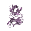
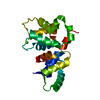
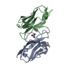
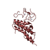
| Title | 2.3 Angstrom resolution structure of dimeric HigBA toxin-antitoxin complex from E. coli | ||||||
 Components Components |
| ||||||
 Keywords Keywords | TOXIN/ANTITOXIN / Toxin-antitoxin complex / HigBA / endoribonuclease / protein-protein complex / PROTEIN BINDING | ||||||
| Function / homology |  Function and homology information Function and homology informationtoxin-antitoxin complex / regulation of growth / core promoter sequence-specific DNA binding / RNA endonuclease activity / regulation of mRNA stability / Hydrolases; Acting on ester bonds / negative regulation of translation / hydrolase activity / regulation of DNA-templated transcription / protein homodimerization activity / RNA binding Similarity search - Function | ||||||
| Biological species |  | ||||||
| Method |  X-RAY DIFFRACTION / SYNCHROTRON / MOLECULAR REPLACEMENT / Resolution: 2.35 Å X-RAY DIFFRACTION / SYNCHROTRON / MOLECULAR REPLACEMENT / Resolution: 2.35 Å | ||||||
 Authors Authors | Jadhav, P. / Sinha, V.K. / Rothweiler, U. / Singh, M. | ||||||
| Funding support |  India, 1items India, 1items
| ||||||
 Citation Citation | Journal: Biochem.J. / Year: 2020 Title: 2.09 angstrom Resolution structure of E. coli HigBA toxin-antitoxin complex reveals an ordered DNA-binding domain and intrinsic dynamics in antitoxin. Authors: Jadhav, P.V. / Sinha, V.K. / Chugh, S. / Kotyada, C. / Bachhav, D. / Singh, R. / Rothweiler, U. / Singh, M. | ||||||
| History |
|
- Structure visualization
| Structure viewer | Molecule: MolmilJmol/JSmol |
|---|
- Downloads & links
-Download
| PDBx/mmCIF format | 6kmq.cif.gz | 94.1 KB | Display | PDBx/mmCIF format |
|---|---|---|---|---|
| PDB format | pdb6kmq.ent.gz | 69.5 KB | Display | PDB format |
| PDBx/mmJSON format | 6kmq.json.gz | Tree view | PDBx/mmJSON format | |
| Others |  Other downloads Other downloads |
-Validation report
| Arichive directory | https://data.pdbj.org/pub/pdb/validation_reports/km/6kmqftp://data.pdbj.org/pub/pdb/validation_reports/km/6kmq | HTTPS FTP |
|---|
-Related structure data
| Related structure data |  6kmlC  5ifgS C: citing same article ( S: Starting model for refinement |
|---|---|
| Similar structure data |


-Links
 PDBj
PDBj


- Assembly
| Deposited unit | 
| ||||||||
|---|---|---|---|---|---|---|---|---|---|
| 1 |
| ||||||||
| Unit cell |
|
-Components
| #1: Protein | Mass: 13676.759 Da / Num. of mol.: 1 Source method: isolated from a genetically manipulated source Source: (gene. exp.) References: UniProt: P64578, Hydrolases; Acting on ester bonds |
|---|---|
| #2: Protein | Mass: 15008.354 Da / Num. of mol.: 1 Source method: isolated from a genetically manipulated source Source: (gene. exp.) |
-Experimental details
-Experiment
| Experiment | Method: X-RAY DIFFRACTION / Number of used crystals: 1 |
|---|
- Sample preparation
| Crystal | Density Matthews: 1.76 Å3/Da / Density % sol: 30.07 % |
|---|---|
| Crystal grow | Temperature: 293 K / Method: vapor diffusion, hanging drop Details: 0.05M MgCl2, 0.2M KCl, 0.1M Tris pH 7.5, 20% PEG 3350 |
-Data collection
| Diffraction | Mean temperature: 100 K / Serial crystal experiment: N |
|---|---|
| Diffraction source | Source: SYNCHROTRON / Site: BESSY  / Beamline: 14.2 / Wavelength: 0.9184 Å / Beamline: 14.2 / Wavelength: 0.9184 Å |
| Detector | Type: DECTRIS PILATUS3 S 6M / Detector: PIXEL / Date: Feb 27, 2018 |
| Radiation | Protocol: SINGLE WAVELENGTH / Monochromatic (M) / Laue (L): M / Scattering type: x-ray |
| Radiation wavelength | Wavelength: 0.9184 Å / Relative weight: 1 |
| Reflection | Resolution: 2.3→28.492 Å / Num. obs: 8815 / % possible obs: 97.5 % / Redundancy: 3.75 % / CC1/2: 0.999 / Net I/σ(I): 13.16 |
| Reflection shell | Resolution: 2.3→2.309 Å / Num. unique obs: 1421 / CC1/2: 0.887 |
- Processing
| Software |
| ||||||||||||||||||||||||||||||||||||||||
|---|---|---|---|---|---|---|---|---|---|---|---|---|---|---|---|---|---|---|---|---|---|---|---|---|---|---|---|---|---|---|---|---|---|---|---|---|---|---|---|---|---|
| Refinement | Method to determine structure: MOLECULAR REPLACEMENT Starting model: 5IFG Resolution: 2.35→28.492 Å / SU ML: 0.29 / Cross valid method: THROUGHOUT / σ(F): 1.36 / Phase error: 38.48
| ||||||||||||||||||||||||||||||||||||||||
| Solvent computation | Shrinkage radii: 0.9 Å / VDW probe radii: 1.11 Å | ||||||||||||||||||||||||||||||||||||||||
| Displacement parameters | Biso max: 121.36 Å2 / Biso mean: 67.778 Å2 / Biso min: 36.22 Å2 | ||||||||||||||||||||||||||||||||||||||||
| Refinement step | Cycle: final / Resolution: 2.35→28.492 Å
| ||||||||||||||||||||||||||||||||||||||||
| LS refinement shell | Refine-ID: X-RAY DIFFRACTION / Rfactor Rfree error: 0 / % reflection obs: 97 %
| ||||||||||||||||||||||||||||||||||||||||
| Refinement TLS params. | Method: refined / Origin x: 8.7668 Å / Origin y: 1.4319 Å / Origin z: 11.0338 Å
| ||||||||||||||||||||||||||||||||||||||||
| Refinement TLS group |
|
