 Movie
Movie Controller


+ Open data

- Basic information
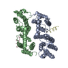
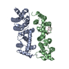
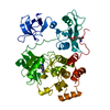
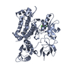
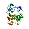
| Entry | Database: PDB / ID: 5ifg | ||||||
|---|---|---|---|---|---|---|---|
| Title | Crystal structure of HigA-HigB complex from E. Coli | ||||||
 Components Components |
| ||||||
 Keywords Keywords | HYDROLASE/ANTITOXIN / mRNA interferase / HYDROLASE-ANTITOXIN complex | ||||||
| Function / homology |  Function and homology information Function and homology informationtoxin-antitoxin complex / regulation of growth / core promoter sequence-specific DNA binding / RNA endonuclease activity / regulation of mRNA stability / Hydrolases; Acting on ester bonds / negative regulation of translation / hydrolase activity / regulation of DNA-templated transcription / protein homodimerization activity / RNA binding Similarity search - Function | ||||||
| Biological species |  | ||||||
| Method |  X-RAY DIFFRACTION / SYNCHROTRON / SAD / Resolution: 2.702 Å X-RAY DIFFRACTION / SYNCHROTRON / SAD / Resolution: 2.702 Å | ||||||
 Authors Authors | Yang, J.S. / Zhou, K. / Gao, z.Q. / Liu, Q.S. / Dong, Y.H. | ||||||
 Citation Citation | Journal: Biochem. Biophys. Res. Commun. / Year: 2016 Title: Structural insight into the E. coli HigBA complex Authors: Yang, J. / Zhou, K. / Liu, P. / Dong, Y. / Gao, Z. / Zhang, J. / Liu, Q. | ||||||
| History |
|
- Structure visualization
| Structure viewer | Molecule: MolmilJmol/JSmol |
|---|
- Downloads & links
-Download
| PDBx/mmCIF format | 5ifg.cif.gz | 96.9 KB | Display | PDBx/mmCIF format |
|---|---|---|---|---|
| PDB format | pdb5ifg.ent.gz | 75.3 KB | Display | PDB format |
| PDBx/mmJSON format | 5ifg.json.gz | Tree view | PDBx/mmJSON format | |
| Others |  Other downloads Other downloads |
-Validation report
| Arichive directory | https://data.pdbj.org/pub/pdb/validation_reports/if/5ifgftp://data.pdbj.org/pub/pdb/validation_reports/if/5ifg | HTTPS FTP |
|---|
-Related structure data
| Similar structure data |
|---|


-Links
 PDBj
PDBj


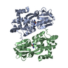
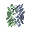
- Assembly
| Deposited unit | 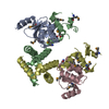
| ||||||||
|---|---|---|---|---|---|---|---|---|---|
| 1 |
| ||||||||
| Unit cell |
|
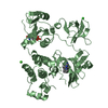
-Components
| #1: Protein | Mass: 12266.822 Da / Num. of mol.: 2 Source method: isolated from a genetically manipulated source Source: (gene. exp.) Strain: K12 / Gene: higB, ygjN, b3083, JW3054 / Production host: References: UniProt: P64578, Hydrolases; Acting on ester bonds #2: Protein | Mass: 15242.830 Da / Num. of mol.: 2 Source method: isolated from a genetically manipulated source Source: (gene. exp.) Strain: K12 / Gene: higA, ygjM, b3082, JW3053 / Production host: Has protein modification | Y | |
|---|
-Experimental details
-Experiment
| Experiment | Method: X-RAY DIFFRACTION / Number of used crystals: 1 |
|---|
- Sample preparation
| Crystal | Density Matthews: 2.88 Å3/Da / Density % sol: 61.46 % |
|---|---|
| Crystal grow | Temperature: 293 K / Method: vapor diffusion, sitting drop / pH: 5.3 Details: 0.2M (NH4)2SO4, 0.1M Bis-Tris pH5.3, 20%(w/v) PEG3350, 0.01M KCl |
-Data collection
| Diffraction | Mean temperature: 100 K |
|---|---|
| Diffraction source | Source: SYNCHROTRON / Site: BSRF  / Beamline: 3W1A / Wavelength: 0.9792 Å / Beamline: 3W1A / Wavelength: 0.9792 Å |
| Detector | Type: MAR CCD 165 mm / Detector: CCD / Date: Jun 3, 2014 |
| Radiation | Protocol: SINGLE WAVELENGTH / Monochromatic (M) / Laue (L): M / Scattering type: x-ray |
| Radiation wavelength | Wavelength: 0.9792 Å / Relative weight: 1 |
| Reflection twin | Operator: -h,-k,l / Fraction: 0.33 |
| Reflection | Resolution: 2.7→50 Å / Num. obs: 35954 / % possible obs: 99.2 % / Redundancy: 17.4 % / Rmerge(I) obs: 0.105 / Net I/σ(I): 65.78 |
| Reflection shell | Resolution: 2.7→2.75 Å / Redundancy: 20.9 % / Rmerge(I) obs: 0.519 / Mean I/σ(I) obs: 14.67 / % possible all: 100 |
- Processing
| Software |
| |||||||||||||||||||||||||||||||||||||||||||||||||||||||||||||||||||||||||||||||||||||||||||
|---|---|---|---|---|---|---|---|---|---|---|---|---|---|---|---|---|---|---|---|---|---|---|---|---|---|---|---|---|---|---|---|---|---|---|---|---|---|---|---|---|---|---|---|---|---|---|---|---|---|---|---|---|---|---|---|---|---|---|---|---|---|---|---|---|---|---|---|---|---|---|---|---|---|---|---|---|---|---|---|---|---|---|---|---|---|---|---|---|---|---|---|---|
| Refinement | Method to determine structure: SAD / Resolution: 2.702→44.928 Å / Cross valid method: FREE R-VALUE / σ(F): 1.34 / Phase error: 31.79 / Stereochemistry target values: TWIN_LSQ_F Details: THE STRUCTURE FACTOR FILE CONTAINS FRIEDEL PAIRS IN F_PLUS/MINUS AND I_PLUS/MINUS COLUMNS
| |||||||||||||||||||||||||||||||||||||||||||||||||||||||||||||||||||||||||||||||||||||||||||
| Solvent computation | Shrinkage radii: 0.9 Å / VDW probe radii: 1.11 Å | |||||||||||||||||||||||||||||||||||||||||||||||||||||||||||||||||||||||||||||||||||||||||||
| Refinement step | Cycle: LAST / Resolution: 2.702→44.928 Å
| |||||||||||||||||||||||||||||||||||||||||||||||||||||||||||||||||||||||||||||||||||||||||||
| Refine LS restraints |
| |||||||||||||||||||||||||||||||||||||||||||||||||||||||||||||||||||||||||||||||||||||||||||
| LS refinement shell |
|
