 Movie
Movie Controller


+ Open data

- Basic information
| Entry | Database: PDB / ID: 6km8 | ||||||
|---|---|---|---|---|---|---|---|
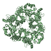
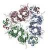
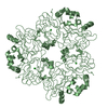
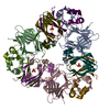
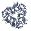
| Title | Crystal Structure of Momordica charantia 7S globulin | ||||||
 Components Components | 7S globulin | ||||||
 Keywords Keywords | PLANT PROTEIN / 7S globulin bi-cupin fold / Glycosylated / Copper-ion | ||||||
| Function / homology | Jelly Rolls / Jelly Rolls / Sandwich / Mainly Beta / ACETATE ION / COPPER (II) ION Function and homology information Function and homology information | ||||||
| Biological species |  Momordica charantia (bitter melon) Momordica charantia (bitter melon) | ||||||
| Method |  X-RAY DIFFRACTION / MOLECULAR REPLACEMENT / Resolution: 3.099 Å X-RAY DIFFRACTION / MOLECULAR REPLACEMENT / Resolution: 3.099 Å | ||||||
 Authors Authors | Kesari, P. / Pratap, S. / Dhankhar, P. / Dalal, V. / Kumar, P. | ||||||
 Citation Citation | Journal: Sci Rep / Year: 2020 Title: Structural characterization and in-silico analysis of Momordica charantia 7S globulin for stability and ACE inhibition. Authors: Kesari, P. / Pratap, S. / Dhankhar, P. / Dalal, V. / Mishra, M. / Singh, P.K. / Chauhan, H. / Kumar, P. | ||||||
| History |
|
- Structure visualization
| Structure viewer | Molecule: MolmilJmol/JSmol |
|---|
- Downloads & links
-Download
| PDBx/mmCIF format | 6km8.cif.gz | 83.8 KB | Display | PDBx/mmCIF format |
|---|---|---|---|---|
| PDB format | pdb6km8.ent.gz | 60.3 KB | Display | PDB format |
| PDBx/mmJSON format | 6km8.json.gz | Tree view | PDBx/mmJSON format | |
| Others |  Other downloads Other downloads |
-Validation report
| Arichive directory | https://data.pdbj.org/pub/pdb/validation_reports/km/6km8ftp://data.pdbj.org/pub/pdb/validation_reports/km/6km8 | HTTPS FTP |
|---|
-Related structure data
| Related structure data |  5e1rS S: Starting model for refinement |
|---|---|
| Similar structure data |


-Links
 PDBj
PDBj- Assembly
| Deposited unit | 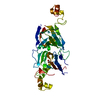
| ||||||||
|---|---|---|---|---|---|---|---|---|---|
| 1 | 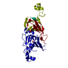
| ||||||||
| Unit cell |
|
-Components
| #1: Protein | Mass: 42996.395 Da / Num. of mol.: 1 / Source method: isolated from a natural source / Source: (natural) Momordica charantia (bitter melon) |
|---|---|
| #2: Chemical | ChemComp-CU /   Mass: 63.546 Da / Num. of mol.: 1 / Source method: obtained synthetically / Formula: Cu Mass: 63.546 Da / Num. of mol.: 1 / Source method: obtained synthetically / Formula: Cu |
| #3: Chemical | ChemComp-ACT /   Mass: 59.044 Da / Num. of mol.: 1 / Source method: obtained synthetically / Formula: C2H3O2 Mass: 59.044 Da / Num. of mol.: 1 / Source method: obtained synthetically / Formula: C2H3O2 |
| #4: Water | ChemComp-HOH /  Mass: 18.015 Da / Num. of mol.: 7 / Source method: isolated from a natural source / Formula: H2O Mass: 18.015 Da / Num. of mol.: 7 / Source method: isolated from a natural source / Formula: H2O |
| Has ligand of interest | N |
-Experimental details
-Experiment
| Experiment | Method: X-RAY DIFFRACTION / Number of used crystals: 1 |
|---|
- Sample preparation
| Crystal | Density Matthews: 1.89 Å3/Da / Density % sol: 35.05 % |
|---|---|
| Crystal grow | Temperature: 293 K / Method: vapor diffusion, sitting drop / pH: 4.6 Details: 0.1 M Sodium acetate trihydrate pH 4.6, 2M Sodium chloride |
-Data collection
| Diffraction | Mean temperature: 100 K / Serial crystal experiment: N |
|---|---|
| Diffraction source | Source: ROTATING ANODE / Type: BRUKER AXS MICROSTAR-H / Wavelength: 1.5418 Å |
| Detector | Type: MARRESEARCH / Detector: CCD / Date: Nov 14, 2011 |
| Radiation | Protocol: SINGLE WAVELENGTH / Monochromatic (M) / Laue (L): M / Scattering type: x-ray |
| Radiation wavelength | Wavelength: 1.5418 Å / Relative weight: 1 |
| Reflection | Resolution: 3.099→50 Å / Num. obs: 5314 / % possible obs: 89.52 % / Redundancy: 2.8 % / Biso Wilson estimate: 53.8 Å2 / Rsym value: 0.12 / Net I/σ(I): 11.17 |
| Reflection shell | Resolution: 3.1→3.15 Å / Redundancy: 2.1 % / Mean I/σ(I) obs: 2.29 / Num. unique obs: 397 / Rsym value: 0.407 / % possible all: 66.44 |
- Processing
| Software |
| ||||||||||||||||||||||||
|---|---|---|---|---|---|---|---|---|---|---|---|---|---|---|---|---|---|---|---|---|---|---|---|---|---|
| Refinement | Method to determine structure: MOLECULAR REPLACEMENT Starting model: 5E1R Resolution: 3.099→45.383 Å / SU ML: 0.45 / Cross valid method: THROUGHOUT / σ(F): 1.35 / Phase error: 33.8
| ||||||||||||||||||||||||
| Solvent computation | Shrinkage radii: 0.9 Å / VDW probe radii: 1.11 Å | ||||||||||||||||||||||||
| Displacement parameters | Biso max: 94.91 Å2 / Biso mean: 53.54 Å2 / Biso min: 12.63 Å2 | ||||||||||||||||||||||||
| Refinement step | Cycle: final / Resolution: 3.099→45.383 Å
| ||||||||||||||||||||||||
| Refine LS restraints |
| ||||||||||||||||||||||||
| LS refinement shell | Resolution: 3.1→3.15 Å / Rfactor Rfree error: 0
|
