 Movie
Movie Controller


+ Open data

- Basic information
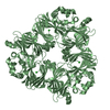
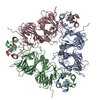
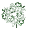
| Entry | Database: PDB / ID: 1fxz | ||||||
|---|---|---|---|---|---|---|---|
| Title | CRYSTAL STRUCTURE OF SOYBEAN PROGLYCININ A1AB1B HOMOTRIMER | ||||||
 Components Components | GLYCININ G1 | ||||||
 Keywords Keywords | PLANT PROTEIN / Proglycinin / Glycinin / Legumin / Seed Storage Protein | ||||||
| Function / homology |  Function and homology information Function and homology informationprotein storage vacuole / nutrient reservoir activity / endoplasmic reticulum Similarity search - Function | ||||||
| Biological species |  | ||||||
| Method |  X-RAY DIFFRACTION / Resolution: 2.8 Å X-RAY DIFFRACTION / Resolution: 2.8 Å | ||||||
 Authors Authors | Adachi, M. / Takenaka, Y. / Gidamis, A.B. / Mikami, B. / Utsumi, S. | ||||||
 Citation Citation | Journal: J.Mol.Biol. / Year: 2001 Title: Crystal structure of soybean proglycinin A1aB1b homotrimer. Authors: Adachi, M. / Takenaka, Y. / Gidamis, A.B. / Mikami, B. / Utsumi, S. #1: Journal: J.Mol.Biol. / Year: 1993Title: Crystallization and preliminary X-ray Crystallographic analysis of the soybean proglycinin expressed in Escherichia coli. Authors: Utsumi, S. / Gidamis, A.B. / Mikami, B. / Kito, M. #2: Journal: Gene / Year: 1988Title: Signal sequence of preproglycinin affects production of the expressed protein in Escherichia coli. Authors: Utsumi, S. / Kim, C.S. / Sato, T. / Kito, M. #3: Journal: J.Agric.Food Chem. / Year: 1987Title: An alternate cDNA encoding glycinin A1aBx subunit Authors: Utsumi, S. / Kohno, T. / Mori, T. / Kito, M. | ||||||
| History |
|
- Structure visualization
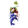
| Structure viewer | Molecule: MolmilJmol/JSmol |
|---|
- Downloads & links
-Download
| PDBx/mmCIF format | 1fxz.cif.gz | 225.3 KB | Display | PDBx/mmCIF format |
|---|---|---|---|---|
| PDB format | pdb1fxz.ent.gz | 180.4 KB | Display | PDB format |
| PDBx/mmJSON format | 1fxz.json.gz | Tree view | PDBx/mmJSON format | |
| Others |  Other downloads Other downloads |
-Validation report
| Arichive directory | https://data.pdbj.org/pub/pdb/validation_reports/fx/1fxzftp://data.pdbj.org/pub/pdb/validation_reports/fx/1fxz | HTTPS FTP |
|---|
-Related structure data
| Similar structure data |
|---|


-Links
 PDBj
PDBj- Assembly
| Deposited unit | 
| ||||||||
|---|---|---|---|---|---|---|---|---|---|
| 1 |
| ||||||||
| Unit cell |
| ||||||||
| Details | The biological assembly is a trimer corresponding to that in asymmetric unit. |
-Components
| #1: Protein | Mass: 53682.500 Da / Num. of mol.: 3 Source method: isolated from a genetically manipulated source Source: (gene. exp.)  #2: Water | ChemComp-HOH / |  Mass: 18.015 Da / Num. of mol.: 34 / Source method: isolated from a natural source / Formula: H2O Mass: 18.015 Da / Num. of mol.: 34 / Source method: isolated from a natural source / Formula: H2OHas protein modification | Y | |
|---|
-Experimental details
-Experiment
| Experiment | Method: X-RAY DIFFRACTION / Number of used crystals: 1 |
|---|
- Sample preparation
| Crystal | Density Matthews: 3.04 Å3/Da / Density % sol: 59.59 % | ||||||||||||||||||||||||||||||
|---|---|---|---|---|---|---|---|---|---|---|---|---|---|---|---|---|---|---|---|---|---|---|---|---|---|---|---|---|---|---|---|
| Crystal grow | Temperature: 302 K / Method: dialysis / pH: 7.6 / Details: pH 7.6, Dialysis, temperature 302.0K | ||||||||||||||||||||||||||||||
| Crystal grow | *PLUS Temperature: 281 K / pH: 5.5 / Method: vapor diffusion, hanging drop | ||||||||||||||||||||||||||||||
| Components of the solutions | *PLUS
|
-Data collection
| Diffraction | Mean temperature: 68 K |
|---|---|
| Diffraction source | Source: ROTATING ANODE / Type: MACSCIENCE / Wavelength: 1.5418 |
| Detector | Type: SIEMENS HI-STAR / Detector: AREA DETECTOR / Date: Jan 17, 1997 |
| Radiation | Protocol: SINGLE WAVELENGTH / Monochromatic (M) / Laue (L): M / Scattering type: x-ray |
| Radiation wavelength | Wavelength: 1.5418 Å / Relative weight: 1 |
| Reflection | Highest resolution: 2.8 Å / Num. obs: 142631 / % possible obs: 99.5 % / Observed criterion σ(F): 0 / Redundancy: 3.1 % |
| Reflection | *PLUS Lowest resolution: 9999 Å / Num. obs: 46010 / Num. measured all: 142631 / Rmerge(I) obs: 0.089 |
- Processing
| Software |
| |||||||||||||||||||||
|---|---|---|---|---|---|---|---|---|---|---|---|---|---|---|---|---|---|---|---|---|---|---|
| Refinement | Resolution: 2.8→8 Å / σ(F): 2
| |||||||||||||||||||||
| Refinement step | Cycle: LAST / Resolution: 2.8→8 Å
| |||||||||||||||||||||
| Software | *PLUS Name: X-PLOR / Version: 3.1 / Classification: refinement | |||||||||||||||||||||
| Refinement | *PLUS Highest resolution: 2.8 Å / Lowest resolution: 8 Å / σ(F): 2 / Rfactor obs: 0.199 / Rfactor Rfree: 0.25 | |||||||||||||||||||||
| Solvent computation | *PLUS | |||||||||||||||||||||
| Displacement parameters | *PLUS | |||||||||||||||||||||
| Refine LS restraints | *PLUS
| |||||||||||||||||||||
| LS refinement shell | *PLUS Highest resolution: 2.8 Å / Lowest resolution: 2.92 Å / Rfactor Rfree: 0.341 / Rfactor obs: 0.271 |
