 Movie
Movie Controller

[English] 日本語
 Yorodumi
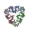
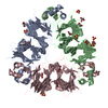
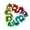
- PDB-3qac: Structure of amaranth 11S proglobulin seed storage protein from A... -
+ Open data

- Basic information
| Entry | Database: PDB / ID: 3qac | ||||||
|---|---|---|---|---|---|---|---|
| Title | Structure of amaranth 11S proglobulin seed storage protein from Amaranthus hypochondriacus L. | ||||||
 Components Components | 11S globulin seed storage protein | ||||||
 Keywords Keywords | PLANT PROTEIN / 11S seed storage protein (globulins) family / seed storage protein | ||||||
| Function / homology |  Function and homology information Function and homology information | ||||||
| Biological species |  Amaranthus hypochondriacus (grain amaranth) Amaranthus hypochondriacus (grain amaranth) | ||||||
| Method |  X-RAY DIFFRACTION / SYNCHROTRON / MOLECULAR REPLACEMENT / Resolution: 2.275 Å X-RAY DIFFRACTION / SYNCHROTRON / MOLECULAR REPLACEMENT / Resolution: 2.275 Å | ||||||
 Authors Authors | Tandang-Silvas, M.R. / Carrazco-Pena, L. / Barba de la Rosa, A.P. / Osuna-Castro, J.A. / Utsumi, S. / Mikami, B. / Maruyama, N. | ||||||
 Citation Citation | Journal: To be Published Title: Structure of amaranth 11S proglobulin, a major seed storage protein from Amaranthus hypochondriacus L. Authors: Tandang-Silvas, M.R. / Carrazco-Pena, L. / Barba de la Rosa, A.P. / Osuna-Castro, J.A. / Utsumi, S. / Mikami, B. / Maruyama, N. #1: Journal: Acta Crystallogr.,Sect.F / Year: 2010 Title: Expression, purification and preliminary crystallization of amaranth 11S proglobulin seed storage protein from Amaranthus hypochondriacus L. Authors: Tandang-Silvas, M.R. / Carrazco-Pena, L. / Barba de la Rosa, A.P. / Osuna-Castro, J.A. / Utsumi, S. / Mikami, B. / Maruyama, N. | ||||||
| History |
|
- Structure visualization
| Structure viewer | Molecule: MolmilJmol/JSmol |
|---|
- Downloads & links
-Download
| PDBx/mmCIF format | 3qac.cif.gz | 89.4 KB | Display | PDBx/mmCIF format |
|---|---|---|---|---|
| PDB format | pdb3qac.ent.gz | 66.2 KB | Display | PDB format |
| PDBx/mmJSON format | 3qac.json.gz | Tree view | PDBx/mmJSON format | |
| Others |  Other downloads Other downloads |
-Validation report
| Arichive directory | https://data.pdbj.org/pub/pdb/validation_reports/qa/3qacftp://data.pdbj.org/pub/pdb/validation_reports/qa/3qac | HTTPS FTP |
|---|
-Related structure data
| Related structure data | 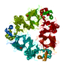 2e9qS S: Starting model for refinement |
|---|---|
| Similar structure data |


-Links
 PDBj
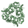
PDBj- Assembly
| Deposited unit | 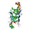
| ||||||||
|---|---|---|---|---|---|---|---|---|---|
| 1 | 
| ||||||||
| Unit cell |
| ||||||||
| Components on special symmetry positions |
|
-Components
| #1: Protein | Mass: 52721.836 Da / Num. of mol.: 1 Source method: isolated from a genetically manipulated source Source: (gene. exp.) Amaranthus hypochondriacus (grain amaranth)Plasmid: pET21d / Production host:  |
|---|---|
| #2: Water | ChemComp-HOH /  Mass: 18.015 Da / Num. of mol.: 100 / Source method: isolated from a natural source / Formula: H2O Mass: 18.015 Da / Num. of mol.: 100 / Source method: isolated from a natural source / Formula: H2O |
| Has protein modification | Y |
-Experimental details
-Experiment
| Experiment | Method: X-RAY DIFFRACTION / Number of used crystals: 1 |
|---|
- Sample preparation
| Crystal | Density Matthews: 1.81 Å3/Da / Density % sol: 32.02 % |
|---|---|
| Crystal grow | Temperature: 293 K / Method: vapor diffusion, hanging drop / pH: 6.6 Details: 16% PEG3350, 200mM ammonium formate, pH 6.6, VAPOR DIFFUSION, HANGING DROP, temperature 293.0K |
-Data collection
| Diffraction | Mean temperature: 100 K |
|---|---|
| Diffraction source | Source: SYNCHROTRON / Site: SPring-8  / Beamline: BL44XU / Wavelength: 0.9 Å / Beamline: BL44XU / Wavelength: 0.9 Å |
| Detector | Type: MARMOSAIC 225 mm CCD / Detector: CCD / Date: Jun 5, 2010 / Details: mirrors |
| Radiation | Protocol: SINGLE WAVELENGTH / Monochromatic (M) / Laue (L): M / Scattering type: x-ray |
| Radiation wavelength | Wavelength: 0.9 Å / Relative weight: 1 |
| Reflection | Resolution: 2.275→50 Å / Num. all: 17239 / Num. obs: 17067 / % possible obs: 99 % / Observed criterion σ(I): 2 / Redundancy: 4.3 % / Biso Wilson estimate: 47.1 Å2 / Rmerge(I) obs: 0.077 / Net I/σ(I): 16.3 |
| Reflection shell | Resolution: 2.3→2.34 Å / Redundancy: 4 % / Rmerge(I) obs: 0.465 / Mean I/σ(I) obs: 4.01 / Num. unique all: 860 / % possible all: 99.3 |
- Processing
| Software |
| |||||||||||||||||||||||||||||||||||||||||||||||||
|---|---|---|---|---|---|---|---|---|---|---|---|---|---|---|---|---|---|---|---|---|---|---|---|---|---|---|---|---|---|---|---|---|---|---|---|---|---|---|---|---|---|---|---|---|---|---|---|---|---|---|
| Refinement | Method to determine structure: MOLECULAR REPLACEMENT Starting model: PDB ENTRY 2E9Q Resolution: 2.275→28.468 Å / Occupancy max: 1 / Occupancy min: 0 / FOM work R set: 0.7811 / SU ML: 0.34 / σ(F): 1.34 / Phase error: 28.43 / Stereochemistry target values: ML
| |||||||||||||||||||||||||||||||||||||||||||||||||
| Solvent computation | Shrinkage radii: 0.9 Å / VDW probe radii: 1.11 Å / Solvent model: FLAT BULK SOLVENT MODEL / Bsol: 42.331 Å2 / ksol: 0.328 e/Å3 | |||||||||||||||||||||||||||||||||||||||||||||||||
| Displacement parameters | Biso max: 97.18 Å2 / Biso mean: 49.2937 Å2 / Biso min: 25.93 Å2
| |||||||||||||||||||||||||||||||||||||||||||||||||
| Refinement step | Cycle: LAST / Resolution: 2.275→28.468 Å
| |||||||||||||||||||||||||||||||||||||||||||||||||
| Refine LS restraints |
| |||||||||||||||||||||||||||||||||||||||||||||||||
| LS refinement shell | Refine-ID: X-RAY DIFFRACTION / Total num. of bins used: 6
|
