Movie
Movie Controller


+ Open data

- Basic information
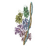
| Entry | Database: PDB / ID: 6klq | ||||||
|---|---|---|---|---|---|---|---|
| Title | Tropomyosin of cardiac thin filament in high-calcium state | ||||||
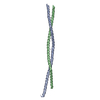
 Components Components | Tropomyosin | ||||||
 Keywords Keywords | CONTRACTILE PROTEIN / Cardiac thin filament | ||||||
| Biological species |  | ||||||
| Method | ELECTRON MICROSCOPY / single particle reconstruction / cryo EM / Resolution: 7.7 Å | ||||||
 Authors Authors | Oda, T. / Yanagisawa, H. / Wakabayashi, T. | ||||||
 Citation Citation | Journal: J Struct Biol / Year: 2020 Title: Cryo-EM structures of cardiac thin filaments reveal the 3D architecture of troponin. Authors: Toshiyuki Oda / Haruaki Yanagisawa / Takeyuki Wakabayashi /  Abstract: Troponin is an essential component of striated muscle and it regulates the sliding of actomyosin system in a calcium-dependent manner. Despite its importance, the structure of troponin has been ...Troponin is an essential component of striated muscle and it regulates the sliding of actomyosin system in a calcium-dependent manner. Despite its importance, the structure of troponin has been elusive due to its high structural heterogeneity. In this study, we analyzed the 3D structures of murine cardiac thin filaments using a cryo-electron microscope equipped with a Volta phase plate (VPP). Contrast enhancement by a VPP enabled us to reconstruct the entire repeat of the thin filament. We determined the orientation of troponin relative to F-actin and tropomyosin, and characterized the interactions between troponin and tropomyosin. This study provides a structural basis for understanding the molecular mechanism of actomyosin system. | ||||||
| History |
|
- Structure visualization
| Movie |
 Movie viewer Movie viewer |
|---|---|
| Structure viewer | Molecule: MolmilJmol/JSmol |
- Downloads & links
-Download
| PDBx/mmCIF format | 6klq.cif.gz | 33.9 KB | Display | PDBx/mmCIF format |
|---|---|---|---|---|
| PDB format | pdb6klq.ent.gz | 26.9 KB | Display | PDB format |
| PDBx/mmJSON format | 6klq.json.gz | Tree view | PDBx/mmJSON format | |
| Others |  Other downloads Other downloads |
-Validation report
| Arichive directory | https://data.pdbj.org/pub/pdb/validation_reports/kl/6klqftp://data.pdbj.org/pub/pdb/validation_reports/kl/6klq | HTTPS FTP |
|---|
-Related structure data
| Related structure data |  0715MC 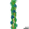 0711C 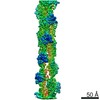 0712C 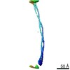 0714C  0717C  0718C 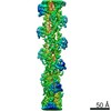 0796C  0797C 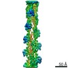 0798C 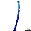 0799C 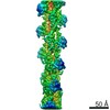 0802C  0804C 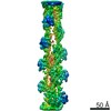 0805C 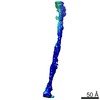 0806C 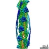 0807C 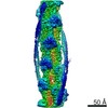 0808C  6kllC 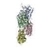 6klnC  6klpC  6kltC  6kluC C: citing same article ( M: map data used to model this data |
|---|---|
| Similar structure data |

-Links
 PDBj
PDBj- Assembly
| Deposited unit | 
|
|---|---|
| 1 |
|
-Components
| #1: Protein | Mass: 11507.176 Da / Num. of mol.: 2 / Source method: isolated from a natural source / Source: (natural) Sequence details | Protein used in this study is tropomyosin 1alpha. Sequence details can be found at https://www.ncbi. ...Protein used in this study is tropomyosin 1alpha. Sequence details can be found at https://www.ncbi.nlm.nih.gov/gene/22003. | |
|---|
-Experimental details
-Experiment
| Experiment | Method: ELECTRON MICROSCOPY |
|---|---|
| EM experiment | Aggregation state: FILAMENT / 3D reconstruction method: single particle reconstruction |
- Sample preparation
| Component | Name: Cardiac thin filament / Type: COMPLEX / Entity ID: all / Source: NATURAL |
|---|---|
| Source (natural) | Organism: |
| Buffer solution | pH: 7.2 |
| Specimen | Conc.: 0.1 mg/ml / Embedding applied: NO / Shadowing applied: NO / Staining applied: NO / Vitrification applied: YES |
| Specimen support | Grid material: COPPER |
| Vitrification | Cryogen name: ETHANE |
- Electron microscopy imaging
| Experimental equipment |  Model: Titan Krios / Image courtesy: FEI Company |
|---|---|
| Microscopy | Model: FEI TITAN KRIOS |
| Electron gun | Electron source:  FIELD EMISSION GUN / Accelerating voltage: 300 kV / Illumination mode: FLOOD BEAM FIELD EMISSION GUN / Accelerating voltage: 300 kV / Illumination mode: FLOOD BEAM |
| Electron lens | Mode: BRIGHT FIELD |
| Image recording | Average exposure time: 5.5 sec. / Electron dose: 60 e/Å2 / Film or detector model: GATAN K3 (6k x 4k) / Num. of real images: 8460 |
- Processing
| EM software |
| ||||||||||||||||||||||||||||||||||||
|---|---|---|---|---|---|---|---|---|---|---|---|---|---|---|---|---|---|---|---|---|---|---|---|---|---|---|---|---|---|---|---|---|---|---|---|---|---|
| CTF correction | Type: PHASE FLIPPING AND AMPLITUDE CORRECTION | ||||||||||||||||||||||||||||||||||||
| Symmetry | Point symmetry: C1 (asymmetric) | ||||||||||||||||||||||||||||||||||||
| 3D reconstruction | Resolution: 7.7 Å / Resolution method: FSC 0.5 CUT-OFF / Num. of particles: 328766 / Symmetry type: POINT | ||||||||||||||||||||||||||||||||||||
| Atomic model building | PDB-ID: 3J8A Accession code: 3J8A / Source name: PDB / Type: experimental model |