 Movie
Movie Controller


+ Open data

- Basic information
| Entry | Database: PDB / ID: 6jl9 | ||||||
|---|---|---|---|---|---|---|---|
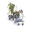
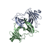
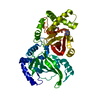
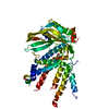
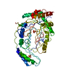
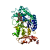
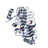
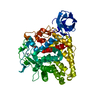
| Title | Crystal structure of a frog ependymin related protein | ||||||
 Components Components | Ependymin-related 1 | ||||||
 Keywords Keywords | UNKNOWN FUNCTION / Ependymin | ||||||
| Function / homology | Ependymin / Ependymin / Ependymins / lysosomal lumen / cell-matrix adhesion / lysosome / calcium ion binding / extracellular region / Mammalian ependymin-related protein 1 Function and homology information Function and homology information | ||||||
| Biological species | |||||||
| Method |  X-RAY DIFFRACTION / SYNCHROTRON / SAD / Resolution: 2 Å X-RAY DIFFRACTION / SYNCHROTRON / SAD / Resolution: 2 Å | ||||||
 Authors Authors | Park, S.Y. | ||||||
| Funding support |  Korea, Republic Of, 1items Korea, Republic Of, 1items
| ||||||
 Citation Citation | Journal: Iucrj / Year: 2019 Title: Structures of three ependymin-related proteins suggest their function as a hydrophobic molecule binder. Authors: Park, J.K. / Kim, K.Y. / Sim, Y.W. / Kim, Y.I. / Kim, J.K. / Lee, C. / Han, J. / Kim, C.U. / Lee, J.E. / Park, S. | ||||||
| History |
|
- Structure visualization
| Structure viewer | Molecule: MolmilJmol/JSmol |
|---|
- Downloads & links
-Download
| PDBx/mmCIF format | 6jl9.cif.gz | 98.2 KB | Display | PDBx/mmCIF format |
|---|---|---|---|---|
| PDB format | pdb6jl9.ent.gz | 74.1 KB | Display | PDB format |
| PDBx/mmJSON format | 6jl9.json.gz | Tree view | PDBx/mmJSON format | |
| Others |  Other downloads Other downloads |
-Validation report
| Arichive directory | https://data.pdbj.org/pub/pdb/validation_reports/jl/6jl9ftp://data.pdbj.org/pub/pdb/validation_reports/jl/6jl9 | HTTPS FTP |
|---|
-Related structure data


-Links
 PDBj
PDBj- Assembly
| Deposited unit | 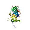
| ||||||||
|---|---|---|---|---|---|---|---|---|---|
| 1 | 
| ||||||||
| Unit cell |
|
-Components
| #1: Protein | Mass: 22836.588 Da / Num. of mol.: 1 Source method: isolated from a genetically manipulated source Source: (gene. exp.) Gene: epdr1 / Production host:   Spodoptera frugiperda (fall armyworm) / References: UniProt: F6VRB7 Spodoptera frugiperda (fall armyworm) / References: UniProt: F6VRB7 |
|---|---|
| #2: Chemical | ChemComp-CA /   Mass: 40.078 Da / Num. of mol.: 1 / Source method: obtained synthetically / Formula: Ca Mass: 40.078 Da / Num. of mol.: 1 / Source method: obtained synthetically / Formula: Ca |
| #3: Water | ChemComp-HOH /  Mass: 18.015 Da / Num. of mol.: 55 / Source method: isolated from a natural source / Formula: H2O Mass: 18.015 Da / Num. of mol.: 55 / Source method: isolated from a natural source / Formula: H2O |
| Has protein modification | Y |
-Experimental details
-Experiment
| Experiment | Method: X-RAY DIFFRACTION / Number of used crystals: 1 |
|---|
- Sample preparation
| Crystal | Density Matthews: 2.8 Å3/Da / Density % sol: 56.02 % |
|---|---|
| Crystal grow | Temperature: 298 K / Method: vapor diffusion, hanging drop Details: 0.2M Calcium acetate hydrate, 0.1M Sodium cacodylate trihydrate pH 6.5, 18%(w/v) Polyethylene glycol 8,000 |
-Data collection
| Diffraction | Mean temperature: 100 K / Serial crystal experiment: N |
|---|---|
| Diffraction source | Source: SYNCHROTRON / Site: PAL/PLS / Beamline: 7A (6B, 6C1) / Wavelength: 1.54 Å |
| Detector | Type: ADSC QUANTUM 270 / Detector: CCD / Date: Jul 25, 2018 |
| Radiation | Protocol: SINGLE WAVELENGTH / Monochromatic (M) / Laue (L): M / Scattering type: x-ray |
| Radiation wavelength | Wavelength: 1.54 Å / Relative weight: 1 |
| Reflection | Resolution: 2→53.01 Å / Num. obs: 18642 / % possible obs: 99.7 % / Redundancy: 19.8 % / Net I/σ(I): 70.5 |
| Reflection shell | Resolution: 2→2.03 Å |
- Processing
| Software |
| ||||||||||||||||||||||||||||||||||||||||||||||||||||||||||||||||||||||||||||||||||||||||||||||||||||||||||||||||||||||||||||||||||||||||||||||||||||||||||||||||||||||||||||||||||||||
|---|---|---|---|---|---|---|---|---|---|---|---|---|---|---|---|---|---|---|---|---|---|---|---|---|---|---|---|---|---|---|---|---|---|---|---|---|---|---|---|---|---|---|---|---|---|---|---|---|---|---|---|---|---|---|---|---|---|---|---|---|---|---|---|---|---|---|---|---|---|---|---|---|---|---|---|---|---|---|---|---|---|---|---|---|---|---|---|---|---|---|---|---|---|---|---|---|---|---|---|---|---|---|---|---|---|---|---|---|---|---|---|---|---|---|---|---|---|---|---|---|---|---|---|---|---|---|---|---|---|---|---|---|---|---|---|---|---|---|---|---|---|---|---|---|---|---|---|---|---|---|---|---|---|---|---|---|---|---|---|---|---|---|---|---|---|---|---|---|---|---|---|---|---|---|---|---|---|---|---|---|---|---|---|
| Refinement | Method to determine structure: SAD / Resolution: 2→53.01 Å / Cor.coef. Fo:Fc: 0.956 / Cor.coef. Fo:Fc free: 0.903 / SU B: 9.651 / SU ML: 0.122 / Cross valid method: THROUGHOUT / ESU R: 0.287 / ESU R Free: 0.171 / Details: HYDROGENS HAVE BEEN ADDED IN THE RIDING POSITIONS
| ||||||||||||||||||||||||||||||||||||||||||||||||||||||||||||||||||||||||||||||||||||||||||||||||||||||||||||||||||||||||||||||||||||||||||||||||||||||||||||||||||||||||||||||||||||||
| Solvent computation | Ion probe radii: 0.8 Å / Shrinkage radii: 0.8 Å / VDW probe radii: 1.2 Å | ||||||||||||||||||||||||||||||||||||||||||||||||||||||||||||||||||||||||||||||||||||||||||||||||||||||||||||||||||||||||||||||||||||||||||||||||||||||||||||||||||||||||||||||||||||||
| Displacement parameters | Biso mean: 55.467 Å2
| ||||||||||||||||||||||||||||||||||||||||||||||||||||||||||||||||||||||||||||||||||||||||||||||||||||||||||||||||||||||||||||||||||||||||||||||||||||||||||||||||||||||||||||||||||||||
| Refinement step | Cycle: 1 / Resolution: 2→53.01 Å
| ||||||||||||||||||||||||||||||||||||||||||||||||||||||||||||||||||||||||||||||||||||||||||||||||||||||||||||||||||||||||||||||||||||||||||||||||||||||||||||||||||||||||||||||||||||||
| Refine LS restraints |
|