 Movie
Movie Controller

[English] 日本語
 Yorodumi
- PDB-3ugs: Crystal structure of a probable undecaprenyl diphosphate synthase... -
+ Open data

- Basic information
| Entry | Database: PDB / ID: 3ugs | ||||||
|---|---|---|---|---|---|---|---|
| Title | Crystal structure of a probable undecaprenyl diphosphate synthase (uppS) from Campylobacter jejuni | ||||||
 Components Components | Undecaprenyl pyrophosphate synthase | ||||||
 Keywords Keywords | TRANSFERASE / NIAID / CSGID / Structural Genomics / Center for Structural Genomics of Infectious Diseases / farnesyl monophosphate | ||||||
| Function / homology |  Function and homology information Function and homology informationditrans,polycis-polyprenyl diphosphate synthase [(2E,6E)-farnesyl diphosphate specific] activity / polyprenol biosynthetic process / Transferases; Transferring alkyl or aryl groups, other than methyl groups / magnesium ion binding Similarity search - Function | ||||||
| Biological species |   Campylobacter jejuni (Campylobacter) Campylobacter jejuni (Campylobacter) | ||||||
| Method |  X-RAY DIFFRACTION / SYNCHROTRON / MOLECULAR REPLACEMENT / Resolution: 2.457 Å X-RAY DIFFRACTION / SYNCHROTRON / MOLECULAR REPLACEMENT / Resolution: 2.457 Å | ||||||
 Authors Authors | Nocek, B. / Gu, M. / Grimshaw, S. / Anderson, W.F. / Joachimiak, A. / Center for Structural Genomics of Infectious Diseases (CSGID) | ||||||
 Citation Citation | Journal: TO BE PUBLISHED Title: Crystal structure of a probable undecaprenyl diphosphate synthase (uppS) from Campylobacter jejuni Authors: Nocek, B. / Gu, M. / Grimshaw, S. / Anderson, W.F. / Joachimiak, A. / Center for Structural Genomics of Infectious Diseases (CSGID) | ||||||
| History |
|
- Structure visualization
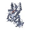
| Structure viewer | Molecule: MolmilJmol/JSmol |
|---|
- Downloads & links
-Download
| PDBx/mmCIF format | 3ugs.cif.gz | 96.1 KB | Display | PDBx/mmCIF format |
|---|---|---|---|---|
| PDB format | pdb3ugs.ent.gz | 72.7 KB | Display | PDB format |
| PDBx/mmJSON format | 3ugs.json.gz | Tree view | PDBx/mmJSON format | |
| Others |  Other downloads Other downloads |
-Validation report
| Arichive directory | https://data.pdbj.org/pub/pdb/validation_reports/ug/3ugsftp://data.pdbj.org/pub/pdb/validation_reports/ug/3ugs | HTTPS FTP |
|---|
-Related structure data
| Related structure data | 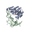 2dr2S S: Starting model for refinement |
|---|---|
| Similar structure data | |
| Other databases |


-Links
 PDBj
PDBj- Assembly
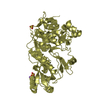
| Deposited unit | 
| ||||||||
|---|---|---|---|---|---|---|---|---|---|
| 1 |
| ||||||||
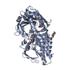
| Unit cell |
|
-Components
| #1: Protein | Mass: 26401.617 Da / Num. of mol.: 2 Source method: isolated from a genetically manipulated source Source: (gene. exp.) Campylobacter jejuni (Campylobacter) / Gene: uppS, Cj0824 / Production host: References: UniProt: Q9PP99, ditrans,polycis-undecaprenyl-diphosphate synthase [(2E,6E)-farnesyl-diphosphate specific] #2: Chemical |   Mass: 302.346 Da / Num. of mol.: 2 / Source method: obtained synthetically / Formula: C15H27O4P Mass: 302.346 Da / Num. of mol.: 2 / Source method: obtained synthetically / Formula: C15H27O4P#3: Water | ChemComp-HOH / |  Mass: 18.015 Da / Num. of mol.: 70 / Source method: isolated from a natural source / Formula: H2O Mass: 18.015 Da / Num. of mol.: 70 / Source method: isolated from a natural source / Formula: H2ONonpolymer details | DURING MODEL BUILDING AND REFINEMENT, AN UNIDENTIFIED LIGAND WAS OBSERVED IN THE ACTIVE SITE AREA. ...DURING MODEL BUILDING AND REFINEMENT | |
|---|
-Experimental details
-Experiment
| Experiment | Method: X-RAY DIFFRACTION / Number of used crystals: 1 |
|---|
- Sample preparation
| Crystal | Density Matthews: 2.29 Å3/Da / Density % sol: 46.39 % |
|---|---|
| Crystal grow | Temperature: 279 K / Method: vapor diffusion, sitting drop / pH: 7 Details: 45% MPD, pH 7.0, VAPOR DIFFUSION, SITTING DROP, temperature 279K |
-Data collection
| Diffraction | Mean temperature: 100 K |
|---|---|
| Diffraction source | Source: SYNCHROTRON / Site: APS  / Beamline: 19-ID / Wavelength: 0.9794 Å / Beamline: 19-ID / Wavelength: 0.9794 Å |
| Detector | Type: ADSC QUANTUM 315r / Detector: CCD / Date: Dec 20, 2009 / Details: mirrors |
| Radiation | Monochromator: double crystal / Protocol: SINGLE WAVELENGTH / Monochromatic (M) / Laue (L): M / Scattering type: x-ray |
| Radiation wavelength | Wavelength: 0.9794 Å / Relative weight: 1 |
| Reflection | Resolution: 2.457→40 Å / Num. all: 18358 / Num. obs: 18362 / % possible obs: 99.8 % / Observed criterion σ(F): 2 / Observed criterion σ(I): 2 / Redundancy: 9.1 % / Rmerge(I) obs: 0.068 / Net I/σ(I): 28 |
| Reflection shell | Resolution: 2.45→2.49 Å / Num. unique all: 908 / % possible all: 100 |
- Processing
| Software |
| ||||||||||||||||||||||||||||||||||||||||||||||||||||||||||||||||||||||||||||||||||||||||||||||||||||||||||||||||||||||||||||||||||||||||||||||||||||||||||||||||||||||||||
|---|---|---|---|---|---|---|---|---|---|---|---|---|---|---|---|---|---|---|---|---|---|---|---|---|---|---|---|---|---|---|---|---|---|---|---|---|---|---|---|---|---|---|---|---|---|---|---|---|---|---|---|---|---|---|---|---|---|---|---|---|---|---|---|---|---|---|---|---|---|---|---|---|---|---|---|---|---|---|---|---|---|---|---|---|---|---|---|---|---|---|---|---|---|---|---|---|---|---|---|---|---|---|---|---|---|---|---|---|---|---|---|---|---|---|---|---|---|---|---|---|---|---|---|---|---|---|---|---|---|---|---|---|---|---|---|---|---|---|---|---|---|---|---|---|---|---|---|---|---|---|---|---|---|---|---|---|---|---|---|---|---|---|---|---|---|---|---|---|---|---|---|
| Refinement | Method to determine structure: MOLECULAR REPLACEMENT Starting model: PDB ENTRY 2DR2 Resolution: 2.457→40 Å / Cor.coef. Fo:Fc: 0.933 / Cor.coef. Fo:Fc free: 0.902 / SU B: 9.259 / SU ML: 0.213 / Cross valid method: THROUGHOUT / σ(F): 2 / ESU R: 0.585 / ESU R Free: 0.3 / Stereochemistry target values: MAXIMUM LIKELIHOOD Details: HYDROGENS HAVE BEEN ADDED IN THE RIDING POSITIONS U VALUES : REFINED INDIVIDUALLY
| ||||||||||||||||||||||||||||||||||||||||||||||||||||||||||||||||||||||||||||||||||||||||||||||||||||||||||||||||||||||||||||||||||||||||||||||||||||||||||||||||||||||||||
| Solvent computation | Ion probe radii: 0.8 Å / Shrinkage radii: 0.8 Å / VDW probe radii: 1.2 Å / Solvent model: MASK | ||||||||||||||||||||||||||||||||||||||||||||||||||||||||||||||||||||||||||||||||||||||||||||||||||||||||||||||||||||||||||||||||||||||||||||||||||||||||||||||||||||||||||
| Displacement parameters | Biso mean: 30.119 Å2
| ||||||||||||||||||||||||||||||||||||||||||||||||||||||||||||||||||||||||||||||||||||||||||||||||||||||||||||||||||||||||||||||||||||||||||||||||||||||||||||||||||||||||||
| Refinement step | Cycle: LAST / Resolution: 2.457→40 Å
| ||||||||||||||||||||||||||||||||||||||||||||||||||||||||||||||||||||||||||||||||||||||||||||||||||||||||||||||||||||||||||||||||||||||||||||||||||||||||||||||||||||||||||
| Refine LS restraints |
| ||||||||||||||||||||||||||||||||||||||||||||||||||||||||||||||||||||||||||||||||||||||||||||||||||||||||||||||||||||||||||||||||||||||||||||||||||||||||||||||||||||||||||
| LS refinement shell | Resolution: 2.457→2.521 Å / Total num. of bins used: 20
|
