 Movie
Movie Controller

[English] 日本語
 Yorodumi
- PDB-4fff: Crystal Structure of Levan Fructotransferase from Arthrobacter ur... -
+ Open data

- Basic information
| Entry | Database: PDB / ID: 4fff | ||||||
|---|---|---|---|---|---|---|---|
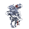
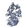
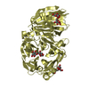
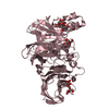
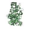
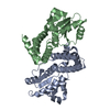
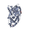
| Title | Crystal Structure of Levan Fructotransferase from Arthrobacter ureafaciens | ||||||
 Components Components | Levan fructotransferase | ||||||
 Keywords Keywords | HYDROLASE / glycoside hydrolase | ||||||
| Function / homology |  Function and homology information Function and homology informationsucrose catabolic process / sucrose alpha-glucosidase activity / transferase activity / cytoplasm Similarity search - Function | ||||||
| Biological species |  Arthrobacter ureafaciens (bacteria) Arthrobacter ureafaciens (bacteria) | ||||||
| Method |  X-RAY DIFFRACTION / SYNCHROTRON / MAD / Resolution: 2.57 Å X-RAY DIFFRACTION / SYNCHROTRON / MAD / Resolution: 2.57 Å | ||||||
 Authors Authors | Park, J. / Rhee, S. | ||||||
 Citation Citation | Journal: J.Biol.Chem. / Year: 2012 Title: Structural and functional basis for substrate specificity and catalysis of levan fructotransferase. Authors: Park, J. / Kim, M.I. / Park, Y.D. / Shin, I. / Cha, J. / Kim, C.H. / Rhee, S. | ||||||
| History |
|
- Structure visualization
| Structure viewer | Molecule: MolmilJmol/JSmol |
|---|
- Downloads & links
-Download
| PDBx/mmCIF format | 4fff.cif.gz | 382.3 KB | Display | PDBx/mmCIF format |
|---|---|---|---|---|
| PDB format | pdb4fff.ent.gz | 311.5 KB | Display | PDB format |
| PDBx/mmJSON format | 4fff.json.gz | Tree view | PDBx/mmJSON format | |
| Others |  Other downloads Other downloads |
-Validation report
| Arichive directory | https://data.pdbj.org/pub/pdb/validation_reports/ff/4fffftp://data.pdbj.org/pub/pdb/validation_reports/ff/4fff | HTTPS FTP |
|---|
-Related structure data


-Links
 PDBj
PDBj

- Assembly
| Deposited unit | 
| ||||||||
|---|---|---|---|---|---|---|---|---|---|
| 1 | 
| ||||||||
| 2 | 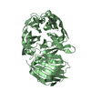
| ||||||||
| 3 | 
| ||||||||
| 4 | 
| ||||||||
| Unit cell |
|
-Components
| #1: Protein | Mass: 54014.406 Da / Num. of mol.: 4 / Fragment: UNP residues 41-521 Source method: isolated from a genetically manipulated source Source: (gene. exp.) Arthrobacter ureafaciens (bacteria) / Gene: lftA / Production host: #2: Water | ChemComp-HOH / |  Mass: 18.015 Da / Num. of mol.: 709 / Source method: isolated from a natural source / Formula: H2O Mass: 18.015 Da / Num. of mol.: 709 / Source method: isolated from a natural source / Formula: H2O |
|---|
-Experimental details
-Experiment
| Experiment | Method: X-RAY DIFFRACTION / Number of used crystals: 1 |
|---|
- Sample preparation
| Crystal | Density Matthews: 4.14 Å3/Da / Density % sol: 70.29 % |
|---|---|
| Crystal grow | Temperature: 295 K / Method: vapor diffusion, hanging drop / pH: 5.5 Details: 0.1M Bis-Tris pH5.5, 1% PEG3350, 1M Ammonium sulfate, 3% 2-propanol, VAPOR DIFFUSION, HANGING DROP, temperature 295K |
-Data collection
| Diffraction | Mean temperature: 100 K | ||||||||||||
|---|---|---|---|---|---|---|---|---|---|---|---|---|---|
| Diffraction source | Source: SYNCHROTRON / Site: PAL/PLS  / Beamline: 6C1 / Wavelength: 0.97949,0.97961,0.97181 / Beamline: 6C1 / Wavelength: 0.97949,0.97961,0.97181 | ||||||||||||
| Detector | Type: ADSC QUANTUM 210 / Detector: CCD | ||||||||||||
| Radiation | Protocol: MAD / Monochromatic (M) / Laue (L): M / Scattering type: x-ray | ||||||||||||
| Radiation wavelength |
| ||||||||||||
| Reflection | Resolution: 2.57→50 Å / Num. all: 112528 / Num. obs: 104570 |
- Processing
| Software |
| ||||||||||||||||||||||||||||
|---|---|---|---|---|---|---|---|---|---|---|---|---|---|---|---|---|---|---|---|---|---|---|---|---|---|---|---|---|---|
| Refinement | Method to determine structure: MAD / Resolution: 2.57→50 Å / Occupancy max: 1 / Occupancy min: 1 / σ(F): 7958
| ||||||||||||||||||||||||||||
| Solvent computation | Bsol: 26.966 Å2 | ||||||||||||||||||||||||||||
| Displacement parameters | Biso max: 63.12 Å2 / Biso mean: 25.9512 Å2 / Biso min: 1 Å2
| ||||||||||||||||||||||||||||
| Refinement step | Cycle: LAST / Resolution: 2.57→50 Å
| ||||||||||||||||||||||||||||
| Refine LS restraints |
| ||||||||||||||||||||||||||||
| Xplor file |
|
