 Movie
Movie Controller

[English] 日本語
 Yorodumi
- PDB-6igk: Crystal Structure of human ETB receptor in complex with Endothelin-3 -
+ Open data

- Basic information
| Entry | Database: PDB / ID: 6igk | ||||||
|---|---|---|---|---|---|---|---|
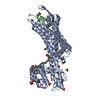
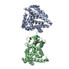
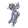
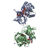
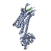
| Title | Crystal Structure of human ETB receptor in complex with Endothelin-3 | ||||||
 Components Components |
| ||||||
 Keywords Keywords | SIGNALING PROTEIN/PROTEIN BINDING / alpha helical / SIGNALING PROTEIN-PROTEIN BINDING complex | ||||||
| Function / homology |  Function and homology information Function and homology informationenteric smooth muscle cell differentiation / response to endothelin / neuroblast migration / negative regulation of neuron maturation / chordate pharynx development / endothelin receptor activity / peptide hormone secretion / endothelin B receptor binding / aldosterone metabolic process / regulation of fever generation ...enteric smooth muscle cell differentiation / response to endothelin / neuroblast migration / negative regulation of neuron maturation / chordate pharynx development / endothelin receptor activity / peptide hormone secretion / endothelin B receptor binding / aldosterone metabolic process / regulation of fever generation / vein smooth muscle contraction / intracellular magnesium ion homeostasis / regulation of developmental pigmentation / positive regulation of penile erection / posterior midgut development / epithelial fluid transport / heparin proteoglycan metabolic process / podocyte differentiation / endothelin receptor signaling pathway / renal sodium excretion / developmental pigmentation / response to sodium phosphate / renin secretion into blood stream / positive regulation of hormone secretion / renal albumin absorption / renal sodium ion absorption / protein transmembrane transport / regulation of pH / vasoconstriction / positive regulation of leukocyte chemotaxis / enteric nervous system development / melanocyte differentiation / peripheral nervous system development / type 1 angiotensin receptor binding / regulation of epithelial cell proliferation / negative regulation of adenylate cyclase activity / : / axon extension / regulation of systemic arterial blood pressure by endothelin / positive regulation of potassium ion transmembrane transport / establishment of endothelial barrier / negative regulation of protein metabolic process / neural crest cell migration / response to pain / positive regulation of urine volume / blood circulation / positive regulation of MAP kinase activity / macrophage chemotaxis / peptide hormone binding / canonical Wnt signaling pathway / regulation of vasoconstriction / viral release from host cell by cytolysis / establishment of localization in cell / positive regulation of heart rate / peptidoglycan catabolic process / neutrophil chemotaxis / positive regulation of mitotic nuclear division / Transcriptional and post-translational regulation of MITF-M expression and activity / potassium ion transmembrane transport / regulation of heart rate / axon guidance / Peptide ligand-binding receptors / calcium-mediated signaling / positive regulation of cell differentiation / hormone activity / vasodilation / intracellular calcium ion homeostasis / cell wall macromolecule catabolic process / calcium ion transmembrane transport / lysozyme / lysozyme activity / cell-cell signaling / nervous system development / positive regulation of cytosolic calcium ion concentration / cellular response to lipopolysaccharide / regulation of gene expression / gene expression / nuclear membrane / G alpha (q) signalling events / phospholipase C-activating G protein-coupled receptor signaling pathway / host cell cytoplasm / positive regulation of canonical NF-kappaB signal transduction / cell population proliferation / cell surface receptor signaling pathway / defense response to bacterium / signaling receptor binding / positive regulation of cell population proliferation / negative regulation of apoptotic process / negative regulation of transcription by RNA polymerase II / signal transduction / : / extracellular region / plasma membrane Similarity search - Function | ||||||
| Biological species |  Homo sapiens (human) Homo sapiens (human) Enterobacteria phage RB59 (virus) Enterobacteria phage RB59 (virus) | ||||||
| Method |  X-RAY DIFFRACTION / SYNCHROTRON / MOLECULAR REPLACEMENT / Resolution: 2 Å X-RAY DIFFRACTION / SYNCHROTRON / MOLECULAR REPLACEMENT / Resolution: 2 Å | ||||||
 Authors Authors | Shihoya, W. / Izume, T. / Inoue, A. / Yamashita, K. / Kadji, F.M.N. / Hirata, K. / Aoki, J. / Nishizawa, T. / Nureki, O. | ||||||
 Citation Citation | Journal: Nat Commun / Year: 2018 Title: Crystal structures of human ETBreceptor provide mechanistic insight into receptor activation and partial activation. Authors: Shihoya, W. / Izume, T. / Inoue, A. / Yamashita, K. / Kadji, F.M.N. / Hirata, K. / Aoki, J. / Nishizawa, T. / Nureki, O. | ||||||
| History |
|
- Structure visualization
| Structure viewer | Molecule: MolmilJmol/JSmol |
|---|
- Downloads & links
-Download
| PDBx/mmCIF format | 6igk.cif.gz | 223.9 KB | Display | PDBx/mmCIF format |
|---|---|---|---|---|
| PDB format | pdb6igk.ent.gz | 178.6 KB | Display | PDB format |
| PDBx/mmJSON format | 6igk.json.gz | Tree view | PDBx/mmJSON format | |
| Others |  Other downloads Other downloads |
-Validation report
| Arichive directory | https://data.pdbj.org/pub/pdb/validation_reports/ig/6igkftp://data.pdbj.org/pub/pdb/validation_reports/ig/6igk | HTTPS FTP |
|---|
-Related structure data
| Related structure data |  6iglC  5glhS S: Starting model for refinement C: citing same article ( |
|---|---|
| Similar structure data | |
| Experimental dataset #1 | Data reference: 10.15785/SBGRID/611 / Data set type: diffraction image data / Metadata reference: 10.15785/SBGRID/611 |


-Links
 PDBj
PDBj


- Assembly
| Deposited unit | 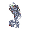
| ||||||||
|---|---|---|---|---|---|---|---|---|---|
| 1 |
| ||||||||
| Unit cell |
|
-Components
| #1: Protein | Mass: 55956.727 Da / Num. of mol.: 1 Mutation: R124Y,D154A,K270A,C1052T,C1095A,S342A,I381A,C396A,C400A,C405A Source method: isolated from a genetically manipulated source Details: Chimera protein of Endothelin receptor type B inserted with Endolysin between residues 303 and 311. Source: (gene. exp.) Homo sapiens (human), (gene. exp.) Enterobacteria phage RB59 (virus)Gene: EDNRB, ETRB, e, RB59_126 / Plasmid: modified pFastBac / Production host:   Spodoptera frugiperda (fall armyworm) / Strain (production host): Sf9 / References: UniProt: P24530, UniProt: A0A097J809, lysozyme Spodoptera frugiperda (fall armyworm) / Strain (production host): Sf9 / References: UniProt: P24530, UniProt: A0A097J809, lysozyme | ||||||
|---|---|---|---|---|---|---|---|
| #2: Protein/peptide | Mass: 2650.100 Da / Num. of mol.: 1 / Source method: obtained synthetically / Source: (synth.) Homo sapiens (human) / References: UniProt: P14138 | ||||||
| #3: Chemical | ChemComp-OLC / (   Mass: 356.540 Da / Num. of mol.: 12 / Source method: obtained synthetically / Formula: C21H40O4 Mass: 356.540 Da / Num. of mol.: 12 / Source method: obtained synthetically / Formula: C21H40O4#4: Chemical | ChemComp-CIT / |   Mass: 192.124 Da / Num. of mol.: 1 / Source method: obtained synthetically / Formula: C6H8O7 Mass: 192.124 Da / Num. of mol.: 1 / Source method: obtained synthetically / Formula: C6H8O7#5: Water | ChemComp-HOH / |  Mass: 18.015 Da / Num. of mol.: 175 / Source method: isolated from a natural source / Formula: H2O Mass: 18.015 Da / Num. of mol.: 175 / Source method: isolated from a natural source / Formula: H2OHas protein modification | Y | |
-Experimental details
-Experiment
| Experiment | Method: X-RAY DIFFRACTION / Number of used crystals: 1 |
|---|
- Sample preparation
| Crystal | Density Matthews: 2.92 Å3/Da / Density % sol: 57.91 % |
|---|---|
| Crystal grow | Temperature: 293 K / Method: lipidic cubic phase / pH: 6 / Details: PEG 500DME, Ammonium citrate tribasic |
-Data collection
| Diffraction | Mean temperature: 100 K | ||||||||||||||||||||||||||||||||||||||||||||||||||||||||||||||||||||||||||||||||
|---|---|---|---|---|---|---|---|---|---|---|---|---|---|---|---|---|---|---|---|---|---|---|---|---|---|---|---|---|---|---|---|---|---|---|---|---|---|---|---|---|---|---|---|---|---|---|---|---|---|---|---|---|---|---|---|---|---|---|---|---|---|---|---|---|---|---|---|---|---|---|---|---|---|---|---|---|---|---|---|---|---|
| Diffraction source | Source: SYNCHROTRON / Site: SPring-8  / Beamline: BL32XU / Wavelength: 1 Å / Beamline: BL32XU / Wavelength: 1 Å | ||||||||||||||||||||||||||||||||||||||||||||||||||||||||||||||||||||||||||||||||
| Detector | Type: DECTRIS EIGER X 9M / Detector: PIXEL / Date: Jul 27, 2016 | ||||||||||||||||||||||||||||||||||||||||||||||||||||||||||||||||||||||||||||||||
| Radiation | Monochromator: Si(111) / Protocol: SINGLE WAVELENGTH / Monochromatic (M) / Laue (L): M / Scattering type: x-ray | ||||||||||||||||||||||||||||||||||||||||||||||||||||||||||||||||||||||||||||||||
| Radiation wavelength | Wavelength: 1 Å / Relative weight: 1 | ||||||||||||||||||||||||||||||||||||||||||||||||||||||||||||||||||||||||||||||||
| Reflection | Resolution: 2→49.599 Å / Num. obs: 46829 / % possible obs: 100 % / Redundancy: 204.625 % / Biso Wilson estimate: 35.54 Å2 / CC1/2: 0.998 / Rmerge(I) obs: 0.858 / Rrim(I) all: 0.86 / Χ2: 1.401 / Net I/σ(I): 20.7 / Num. measured all: 9582405 / Scaling rejects: 4327 | ||||||||||||||||||||||||||||||||||||||||||||||||||||||||||||||||||||||||||||||||
| Reflection shell | Diffraction-ID: 1
|
- Processing
| Software |
| |||||||||||||||||||||||||||||||||||||||||||||||||||||||||||||||||||||||||||||||||||||||||||||||||||||||||||||||||||||||||||||||||||||||||||||||||||||||||||||||||||||||||||||||
|---|---|---|---|---|---|---|---|---|---|---|---|---|---|---|---|---|---|---|---|---|---|---|---|---|---|---|---|---|---|---|---|---|---|---|---|---|---|---|---|---|---|---|---|---|---|---|---|---|---|---|---|---|---|---|---|---|---|---|---|---|---|---|---|---|---|---|---|---|---|---|---|---|---|---|---|---|---|---|---|---|---|---|---|---|---|---|---|---|---|---|---|---|---|---|---|---|---|---|---|---|---|---|---|---|---|---|---|---|---|---|---|---|---|---|---|---|---|---|---|---|---|---|---|---|---|---|---|---|---|---|---|---|---|---|---|---|---|---|---|---|---|---|---|---|---|---|---|---|---|---|---|---|---|---|---|---|---|---|---|---|---|---|---|---|---|---|---|---|---|---|---|---|---|---|---|---|
| Refinement | Method to determine structure: MOLECULAR REPLACEMENT Starting model: 5GLH Resolution: 2→49.599 Å / SU ML: 0.25 / Cross valid method: FREE R-VALUE / σ(F): 1.35 / Phase error: 23.03
| |||||||||||||||||||||||||||||||||||||||||||||||||||||||||||||||||||||||||||||||||||||||||||||||||||||||||||||||||||||||||||||||||||||||||||||||||||||||||||||||||||||||||||||||
| Solvent computation | Shrinkage radii: 0.9 Å / VDW probe radii: 1.11 Å | |||||||||||||||||||||||||||||||||||||||||||||||||||||||||||||||||||||||||||||||||||||||||||||||||||||||||||||||||||||||||||||||||||||||||||||||||||||||||||||||||||||||||||||||
| Displacement parameters | Biso max: 137.61 Å2 / Biso mean: 47.0245 Å2 / Biso min: 20.75 Å2 | |||||||||||||||||||||||||||||||||||||||||||||||||||||||||||||||||||||||||||||||||||||||||||||||||||||||||||||||||||||||||||||||||||||||||||||||||||||||||||||||||||||||||||||||
| Refinement step | Cycle: final / Resolution: 2→49.599 Å
| |||||||||||||||||||||||||||||||||||||||||||||||||||||||||||||||||||||||||||||||||||||||||||||||||||||||||||||||||||||||||||||||||||||||||||||||||||||||||||||||||||||||||||||||
| LS refinement shell | Refine-ID: X-RAY DIFFRACTION / Rfactor Rfree error: 0 / Total num. of bins used: 14 / % reflection obs: 100 %
| |||||||||||||||||||||||||||||||||||||||||||||||||||||||||||||||||||||||||||||||||||||||||||||||||||||||||||||||||||||||||||||||||||||||||||||||||||||||||||||||||||||||||||||||
| Refinement TLS params. | Method: refined / Refine-ID: X-RAY DIFFRACTION
| |||||||||||||||||||||||||||||||||||||||||||||||||||||||||||||||||||||||||||||||||||||||||||||||||||||||||||||||||||||||||||||||||||||||||||||||||||||||||||||||||||||||||||||||
| Refinement TLS group |
|
