 Movie
Movie Controller

[English] 日本語
 Yorodumi
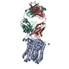
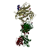
- PDB-6i04: Crystal structure of Sema domain of the Met receptor in complex w... -
+ Open data

- Basic information
| Entry | Database: PDB / ID: 6i04 | ||||||
|---|---|---|---|---|---|---|---|
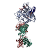
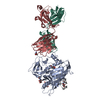
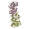
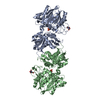
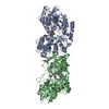
| Title | Crystal structure of Sema domain of the Met receptor in complex with FAB | ||||||
 Components Components |
| ||||||
 Keywords Keywords | IMMUNE SYSTEM / MET / Sema domain | ||||||
| Function / homology |  Function and homology information Function and homology informationhepatocyte growth factor receptor activity / Drug-mediated inhibition of MET activation / MET activates STAT3 / negative regulation of hydrogen peroxide-mediated programmed cell death / MET Receptor Activation / MET interacts with TNS proteins / endothelial cell morphogenesis / semaphorin receptor activity / MET receptor recycling / pancreas development ...hepatocyte growth factor receptor activity / Drug-mediated inhibition of MET activation / MET activates STAT3 / negative regulation of hydrogen peroxide-mediated programmed cell death / MET Receptor Activation / MET interacts with TNS proteins / endothelial cell morphogenesis / semaphorin receptor activity / MET receptor recycling / pancreas development / MET activates PTPN11 / hepatocyte growth factor receptor signaling pathway / MET activates RAP1 and RAC1 / Sema4D mediated inhibition of cell attachment and migration / positive regulation of endothelial cell chemotaxis / MET activates PI3K/AKT signaling / MET activates PTK2 signaling / branching morphogenesis of an epithelial tube / positive chemotaxis / semaphorin-plexin signaling pathway / Regulation of MITF-M-dependent genes involved in cell cycle and proliferation / MET activates RAS signaling / MECP2 regulates neuronal receptors and channels / cell surface receptor protein tyrosine kinase signaling pathway / basal plasma membrane / molecular function activator activity / excitatory postsynaptic potential / liver development / negative regulation of autophagy / InlB-mediated entry of Listeria monocytogenes into host cell / receptor protein-tyrosine kinase / Negative regulation of MET activity / neuron differentiation / Constitutive Signaling by Aberrant PI3K in Cancer / PIP3 activates AKT signaling / PI5P, PP2A and IER3 Regulate PI3K/AKT Signaling / RAF/MAP kinase cascade / protein tyrosine kinase activity / protein phosphatase binding / cell surface receptor signaling pathway / postsynapse / signaling receptor complex / cell surface / positive regulation of transcription by RNA polymerase II / extracellular region / ATP binding / membrane / identical protein binding / plasma membrane Similarity search - Function | ||||||
| Biological species |  Homo sapiens (human) Homo sapiens (human) | ||||||
| Method |  X-RAY DIFFRACTION / SYNCHROTRON / MOLECULAR REPLACEMENT / Resolution: 3.1 Å X-RAY DIFFRACTION / SYNCHROTRON / MOLECULAR REPLACEMENT / Resolution: 3.1 Å | ||||||
 Authors Authors | Casaletto, J.B. / Geddie, M.L. / Abu-Yousif, A.O. / Masson, K. / Fulgham, A. / Boudot, A. / Maiwald, T. / Kearns, J.D. / Kohli, N. / Su, S. ...Casaletto, J.B. / Geddie, M.L. / Abu-Yousif, A.O. / Masson, K. / Fulgham, A. / Boudot, A. / Maiwald, T. / Kearns, J.D. / Kohli, N. / Su, S. / Razlog, M. / Raue, A. / Kalra, A. / Hakansson, M. / Logan, D.T. / Welin, M. / Chattopadhyay, S. / Harms, B.D. / Nielsen, U.B. / Schoeberl, B. / Lugovskoy, A.A. / MacBeath, G. | ||||||
 Citation Citation | Journal: Proc.Natl.Acad.Sci.USA / Year: 2019 Title: MM-131, a bispecific anti-Met/EpCAM mAb, inhibits HGF-dependent and HGF-independent Met signaling through concurrent binding to EpCAM. Authors: Casaletto, J.B. / Geddie, M.L. / Abu-Yousif, A.O. / Masson, K. / Fulgham, A. / Boudot, A. / Maiwald, T. / Kearns, J.D. / Kohli, N. / Su, S. / Razlog, M. / Raue, A. / Kalra, A. / Hakansson, M. ...Authors: Casaletto, J.B. / Geddie, M.L. / Abu-Yousif, A.O. / Masson, K. / Fulgham, A. / Boudot, A. / Maiwald, T. / Kearns, J.D. / Kohli, N. / Su, S. / Razlog, M. / Raue, A. / Kalra, A. / Hakansson, M. / Logan, D.T. / Welin, M. / Chattopadhyay, S. / Harms, B.D. / Nielsen, U.B. / Schoeberl, B. / Lugovskoy, A.A. / MacBeath, G. | ||||||
| History |
|
- Structure visualization
| Structure viewer | Molecule: MolmilJmol/JSmol |
|---|
- Downloads & links
-Download
| PDBx/mmCIF format | 6i04.cif.gz | 349.6 KB | Display | PDBx/mmCIF format |
|---|---|---|---|---|
| PDB format | pdb6i04.ent.gz | 275.3 KB | Display | PDB format |
| PDBx/mmJSON format | 6i04.json.gz | Tree view | PDBx/mmJSON format | |
| Others |  Other downloads Other downloads |
-Validation report
| Arichive directory | https://data.pdbj.org/pub/pdb/validation_reports/i0/6i04ftp://data.pdbj.org/pub/pdb/validation_reports/i0/6i04 | HTTPS FTP |
|---|
-Related structure data
| Related structure data |  6hygC 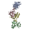 6i07C 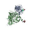 4o3tS  4py7S S: Starting model for refinement C: citing same article ( |
|---|---|
| Similar structure data |


-Links
 PDBj
PDBj


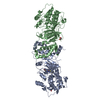
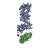
- Assembly
| Deposited unit | 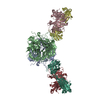
| |||||||||||||||||||||||||||||||||||||||||||||||||||||||||||||||||||||||||||||||||||||||||||||||
|---|---|---|---|---|---|---|---|---|---|---|---|---|---|---|---|---|---|---|---|---|---|---|---|---|---|---|---|---|---|---|---|---|---|---|---|---|---|---|---|---|---|---|---|---|---|---|---|---|---|---|---|---|---|---|---|---|---|---|---|---|---|---|---|---|---|---|---|---|---|---|---|---|---|---|---|---|---|---|---|---|---|---|---|---|---|---|---|---|---|---|---|---|---|---|---|---|
| 1 | 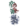
| |||||||||||||||||||||||||||||||||||||||||||||||||||||||||||||||||||||||||||||||||||||||||||||||
| 2 | 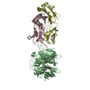
| |||||||||||||||||||||||||||||||||||||||||||||||||||||||||||||||||||||||||||||||||||||||||||||||
| Unit cell |
| |||||||||||||||||||||||||||||||||||||||||||||||||||||||||||||||||||||||||||||||||||||||||||||||
| Noncrystallographic symmetry (NCS) | NCS domain:
NCS domain segments: Component-ID: _ / Refine code: _
NCS ensembles :
|
-Components
| #1: Protein | Mass: 62404.688 Da / Num. of mol.: 2 Source method: isolated from a genetically manipulated source Source: (gene. exp.) Homo sapiens (human) / Gene: MET / Cell line (production host): EXPI293F cells / Production host: Homo sapiens (human)References: UniProt: P08581, receptor protein-tyrosine kinase #2: Antibody | Mass: 24478.488 Da / Num. of mol.: 2 Source method: isolated from a genetically manipulated source Source: (gene. exp.) Homo sapiens (human) / Cell line (production host): EXPI293F cells / Production host: Homo sapiens (human)#3: Antibody | Mass: 23114.676 Da / Num. of mol.: 2 Source method: isolated from a genetically manipulated source Source: (gene. exp.) Homo sapiens (human) / Cell line (production host): EXPI293F cells / Production host: Homo sapiens (human)Has protein modification | Y | |
|---|
-Experimental details
-Experiment
| Experiment | Method: X-RAY DIFFRACTION / Number of used crystals: 1 |
|---|
- Sample preparation
| Crystal | Density Matthews: 2.85 Å3/Da / Density % sol: 56.9 % |
|---|---|
| Crystal grow | Temperature: 293 K / Method: vapor diffusion, sitting drop / Details: 18% w/v PEG 3350, 0.1 M ammonium citrate |
-Data collection
| Diffraction | Mean temperature: 100 K / Serial crystal experiment: N |
|---|---|
| Diffraction source | Source: SYNCHROTRON / Site: Diamond  / Beamline: I02 / Wavelength: 0.9795 Å / Beamline: I02 / Wavelength: 0.9795 Å |
| Detector | Type: DECTRIS PILATUS3 S 6M / Detector: PIXEL / Date: Mar 4, 2016 |
| Radiation | Protocol: SINGLE WAVELENGTH / Monochromatic (M) / Laue (L): M / Scattering type: x-ray |
| Radiation wavelength | Wavelength: 0.9795 Å / Relative weight: 1 |
| Reflection | Resolution: 3.1→29.57 Å / Num. obs: 39528 / % possible obs: 96.9 % / Redundancy: 3.6 % / Biso Wilson estimate: 71 Å2 / CC1/2: 0.987 / Rmerge(I) obs: 0.169 / Net I/σ(I): 5.2 |
| Reflection shell | Resolution: 3.1→3.23 Å / Redundancy: 3.1 % / Rmerge(I) obs: 1.033 / Mean I/σ(I) obs: 1 / Num. unique obs: 3994 / CC1/2: 0.446 / % possible all: 86.1 |
- Processing
| Software |
| ||||||||||||||||||||||||||||||||||||||||||||||||||||||||||||||||||||||||||||||||||||||||||||||||||||||||||||||||||||||||||||||||||||||||||||||||||||||||||||||||||||||||||||||||||||||
|---|---|---|---|---|---|---|---|---|---|---|---|---|---|---|---|---|---|---|---|---|---|---|---|---|---|---|---|---|---|---|---|---|---|---|---|---|---|---|---|---|---|---|---|---|---|---|---|---|---|---|---|---|---|---|---|---|---|---|---|---|---|---|---|---|---|---|---|---|---|---|---|---|---|---|---|---|---|---|---|---|---|---|---|---|---|---|---|---|---|---|---|---|---|---|---|---|---|---|---|---|---|---|---|---|---|---|---|---|---|---|---|---|---|---|---|---|---|---|---|---|---|---|---|---|---|---|---|---|---|---|---|---|---|---|---|---|---|---|---|---|---|---|---|---|---|---|---|---|---|---|---|---|---|---|---|---|---|---|---|---|---|---|---|---|---|---|---|---|---|---|---|---|---|---|---|---|---|---|---|---|---|---|---|
| Refinement | Method to determine structure: MOLECULAR REPLACEMENT Starting model: 4PY7, 4O3T Resolution: 3.1→29.56 Å / Cor.coef. Fo:Fc: 0.94 / Cor.coef. Fo:Fc free: 0.902 / SU B: 29.744 / SU ML: 0.475 / Cross valid method: THROUGHOUT / ESU R Free: 0.497 / Details: HYDROGENS HAVE BEEN ADDED IN THE RIDING POSITIONS
| ||||||||||||||||||||||||||||||||||||||||||||||||||||||||||||||||||||||||||||||||||||||||||||||||||||||||||||||||||||||||||||||||||||||||||||||||||||||||||||||||||||||||||||||||||||||
| Solvent computation | Ion probe radii: 0.8 Å / Shrinkage radii: 0.8 Å / VDW probe radii: 1.2 Å | ||||||||||||||||||||||||||||||||||||||||||||||||||||||||||||||||||||||||||||||||||||||||||||||||||||||||||||||||||||||||||||||||||||||||||||||||||||||||||||||||||||||||||||||||||||||
| Displacement parameters | Biso mean: 82.843 Å2
| ||||||||||||||||||||||||||||||||||||||||||||||||||||||||||||||||||||||||||||||||||||||||||||||||||||||||||||||||||||||||||||||||||||||||||||||||||||||||||||||||||||||||||||||||||||||
| Refinement step | Cycle: 1 / Resolution: 3.1→29.56 Å
| ||||||||||||||||||||||||||||||||||||||||||||||||||||||||||||||||||||||||||||||||||||||||||||||||||||||||||||||||||||||||||||||||||||||||||||||||||||||||||||||||||||||||||||||||||||||
| Refine LS restraints |
|