| 登録情報 | データベース: PDB / ID: 6hyn
|
|---|
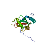
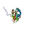
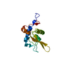
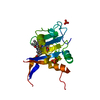
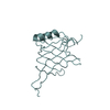
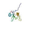
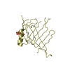
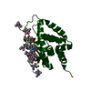
| タイトル | Structure of ATG13 LIR motif bound to GABARAP |
|---|
 要素 要素 | Autophagy-related protein 13,Gamma-aminobutyric acid receptor-associated protein |
|---|
 キーワード キーワード | SIGNALING PROTEIN / Autophagy / ATG8 / LIR |
|---|
| 機能・相同性 |  機能・相同性情報 機能・相同性情報
positive regulation of protein K48-linked ubiquitination / Atg1/ULK1 kinase complex / response to mitochondrial depolarisation / regulation of Rac protein signal transduction / protein localization to phagophore assembly site / phagophore assembly site membrane / GABA receptor binding / piecemeal microautophagy of the nucleus / phosphatidylethanolamine binding / phagophore assembly site ...positive regulation of protein K48-linked ubiquitination / Atg1/ULK1 kinase complex / response to mitochondrial depolarisation / regulation of Rac protein signal transduction / protein localization to phagophore assembly site / phagophore assembly site membrane / GABA receptor binding / piecemeal microautophagy of the nucleus / phosphatidylethanolamine binding / phagophore assembly site / protein kinase regulator activity / TBC/RABGAPs / cellular response to nitrogen starvation / positive regulation of protein targeting to mitochondrion / microtubule associated complex / Macroautophagy / regulation of neurotransmitter receptor localization to postsynaptic specialization membrane / smooth endoplasmic reticulum / autophagosome membrane / extrinsic apoptotic signaling pathway via death domain receptors / axoneme / autophagosome assembly / autophagosome maturation / beta-tubulin binding / protein targeting / mitophagy / sperm midpiece / positive regulation of autophagy / autophagosome / protein serine/threonine kinase activator activity / GABA-ergic synapse / microtubule cytoskeleton organization / positive regulation of proteasomal ubiquitin-dependent protein catabolic process / protein transport / actin cytoskeleton / cytoplasmic vesicle / microtubule binding / chemical synaptic transmission / microtubule / lysosome / Golgi membrane / negative regulation of cell population proliferation / intracellular membrane-bounded organelle / ubiquitin protein ligase binding / endoplasmic reticulum membrane / protein kinase binding / mitochondrion / plasma membrane / cytosol / cytoplasm類似検索 - 分子機能 Autophagy-related protein 13 / HORMA domain superfamily / Autophagy protein Atg8 ubiquitin-like / Autophagy protein Atg8 ubiquitin like / Phosphatidylinositol 3-kinase Catalytic Subunit; Chain A, domain 1 / Ubiquitin-like (UB roll) / Ubiquitin-like domain superfamily / Roll / Alpha Beta類似検索 - ドメイン・相同性 Autophagy-related protein 13 / Gamma-aminobutyric acid receptor-associated protein類似検索 - 構成要素 |
|---|
| 生物種 |  Homo sapiens (ヒト) Homo sapiens (ヒト) |
|---|
| 手法 |  X線回折 / シンクロトロン / 分子置換 / 解像度: 1.14 Å X線回折 / シンクロトロン / 分子置換 / 解像度: 1.14 Å |
|---|
 データ登録者 データ登録者 | Mouilleron, S. / Wirth, M. / Zhang, W. / O'Reilly, N. / Tooze, S. / Johansen, T. / Razi, M. / Nyoni, L. / Joshi, D. |
|---|
 引用 引用 | ジャーナル: Nat Commun / 年: 2019
タイトル: Molecular determinants regulating selective binding of autophagy adapters and receptors to ATG8 proteins.
著者: Wirth, M. / Zhang, W. / Razi, M. / Nyoni, L. / Joshi, D. / O'Reilly, N. / Johansen, T. / Tooze, S.A. / Mouilleron, S. |
|---|
| 履歴 | | 登録 | 2018年10月22日 | 登録サイト: PDBE / 処理サイト: PDBE |
|---|
| 改定 1.0 | 2019年5月8日 | Provider: repository / タイプ: Initial release |
|---|
| 改定 1.1 | 2019年5月15日 | Group: Data collection / Database references
カテゴリ: citation / citation_author / pdbx_database_proc
Item: _citation.journal_volume / _citation.page_first ..._citation.journal_volume / _citation.page_first / _citation.page_last / _citation.pdbx_database_id_PubMed / _citation.title / _citation_author.identifier_ORCID / _citation_author.name |
|---|
| 改定 1.2 | 2024年1月24日 | Group: Data collection / Database references / Refinement description
カテゴリ: chem_comp_atom / chem_comp_bond ...chem_comp_atom / chem_comp_bond / database_2 / pdbx_initial_refinement_model
Item: _database_2.pdbx_DOI / _database_2.pdbx_database_accession |
|---|
|
|---|
 ムービー
ムービー コントローラー


 データを開く
 基本情報
基本情報 構造の表示
構造の表示 ダウンロードとリンク
 その他のダウンロード


 PDBj
PDBj


 集合体
集合体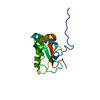

 分子量: 18.015 Da / 分子数: 171 / 由来タイプ: 天然 / 式: H2O
 試料調製
試料調製 / ビームライン: I04-1 / 波長: 0.9159 Å
 解析
解析