- PDB-6hyl: Structure of PCM1 LIR motif bound to GABARAP -
+
Open data
ID or keywords:
Loading...
-
Basic information
Entry
Database: PDB / ID: 6hyl
Title
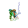
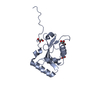
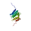
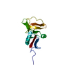
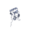
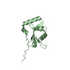
Structure of PCM1 LIR motif bound to GABARAP
Components
Pericentriolar material 1 protein,Gamma-aminobutyric acid receptor-associated protein
Keywords
SIGNALING PROTEIN / Autophagy / ATG8 / LIR
Function / homology
Function and homology information
protein-containing complex localization to centriolar satellite / intraciliary transport involved in cilium assembly / interkinetic nuclear migration / microtubule anchoring / positive regulation of protein K48-linked ubiquitination / microtubule anchoring at centrosome / regulation of Rac protein signal transduction / positive regulation of intracellular protein transport / regulation of protein complex stability / GABA receptor binding ...protein-containing complex localization to centriolar satellite / intraciliary transport involved in cilium assembly / interkinetic nuclear migration / microtubule anchoring / positive regulation of protein K48-linked ubiquitination / microtubule anchoring at centrosome / regulation of Rac protein signal transduction / positive regulation of intracellular protein transport / regulation of protein complex stability / GABA receptor binding / phosphatidylethanolamine binding / non-motile cilium assembly / protein localization to centrosome / TBC/RABGAPs / cellular response to nitrogen starvation / microtubule associated complex / centrosome cycle / Macroautophagy / ciliary transition zone / pericentriolar material / regulation of neurotransmitter receptor localization to postsynaptic specialization membrane / smooth endoplasmic reticulum / autophagosome membrane / cilium assembly / extrinsic apoptotic signaling pathway via death domain receptors / autophagosome maturation / axoneme / autophagosome assembly / beta-tubulin binding / mitophagy / protein targeting / cytoplasmic microtubule organization / Loss of Nlp from mitotic centrosomes / Loss of proteins required for interphase microtubule organization from the centrosome / Recruitment of mitotic centrosome proteins and complexes / Recruitment of NuMA to mitotic centrosomes / Anchoring of the basal body to the plasma membrane / autophagosome / AURKA Activation by TPX2 / centriole / negative regulation of neurogenesis / microtubule cytoskeleton organization / GABA-ergic synapse / centriolar satellite / apical part of cell / Regulation of PLK1 Activity at G2/M Transition / positive regulation of proteasomal ubiquitin-dependent protein catabolic process / protein transport / actin cytoskeleton / cytoplasmic vesicle / sperm midpiece / microtubule binding / molecular adaptor activity / microtubule / chemical synaptic transmission / lysosome / ciliary basal body / Golgi membrane / ubiquitin protein ligase binding / centrosome / protein-containing complex / membrane / identical protein binding / plasma membrane / cytoplasm / cytosol Similarity search - Function
Pericentriolar material 1 protein / Pericentriolar material 1 protein, C-terminal / Pericentriolar material 1 C terminus / Autophagy protein Atg8 ubiquitin-like / Autophagy protein Atg8 ubiquitin like / Phosphatidylinositol 3-kinase Catalytic Subunit; Chain A, domain 1 / Ubiquitin-like (UB roll) / Ubiquitin-like domain superfamily / Roll / Alpha Beta Similarity search - Domain/homology
Gamma-aminobutyric acid receptor-associated protein / Pericentriolar material 1 protein Similarity search - Component
A: Pericentriolar material 1 protein,Gamma-aminobutyric acid receptor-associated protein B: Pericentriolar material 1 protein,Gamma-aminobutyric acid receptor-associated protein
In the structure databanks used in Yorodumi, some data are registered as the other names, "COVID-19 virus" and "2019-nCoV". Here are the details of the virus and the list of structure data.
Jan 31, 2019. EMDB accession codes are about to change! (news from PDBe EMDB page)
EMDB accession codes are about to change! (news from PDBe EMDB page)
The allocation of 4 digits for EMDB accession codes will soon come to an end. Whilst these codes will remain in use, new EMDB accession codes will include an additional digit and will expand incrementally as the available range of codes is exhausted. The current 4-digit format prefixed with “EMD-” (i.e. EMD-XXXX) will advance to a 5-digit format (i.e. EMD-XXXXX), and so on. It is currently estimated that the 4-digit codes will be depleted around Spring 2019, at which point the 5-digit format will come into force.
The EM Navigator/Yorodumi systems omit the EMD- prefix.
Related info.:Q: What is EMD? / ID/Accession-code notation in Yorodumi/EM Navigator
Yorodumi is a browser for structure data from EMDB, PDB, SASBDB, etc.
This page is also the successor to EM Navigator detail page, and also detail information page/front-end page for Omokage search.
The word "yorodu" (or yorozu) is an old Japanese word meaning "ten thousand". "mi" (miru) is to see.
Related info.:EMDB / PDB / SASBDB / Comparison of 3 databanks / Yorodumi Search / Aug 31, 2016. New EM Navigator & Yorodumi / Yorodumi Papers / Jmol/JSmol / Function and homology information / Changes in new EM Navigator and Yorodumi
 Movie
Movie Controller


 Open data
 Basic information
 Components
 Keywords
 Function and homology information
 Homo sapiens (human)
 X-RAY DIFFRACTION /
  Authors
 Citation
 Structure visualization
 Downloads & links
 Other downloads


 PDBj
PDBj


 Assembly
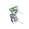

 Mass: 18.015 Da / Num. of mol.: 234 / Source method: isolated from a natural source / Formula: H2O
 Sample preparation
 / Beamline: I04 / Wavelength: 0.9795 Å
 Processing