 Movie
Movie Controller


+ Open data

- Basic information
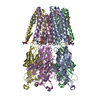
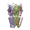
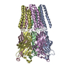
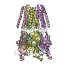
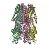
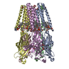
| Entry | Database: PDB / ID: 6hya | ||||||
|---|---|---|---|---|---|---|---|
| Title | THE GLIC PENTAMERIC LIGAND-GATED ION CHANNEL MUTANT Q193L | ||||||
 Components Components | Proton-gated ion channel | ||||||
 Keywords Keywords | MEMBRANE PROTEIN / PENTAMERIC TRANSMEMBRANE CHANNEL / ION CHANNEL. | ||||||
| Function / homology |  Function and homology information Function and homology informationsodium channel activity / potassium channel activity / extracellular ligand-gated monoatomic ion channel activity / transmembrane signaling receptor activity / identical protein binding / plasma membrane Similarity search - Function | ||||||
| Biological species |  Gloeobacter violaceus PCC 7421 (bacteria) Gloeobacter violaceus PCC 7421 (bacteria) | ||||||
| Method |  X-RAY DIFFRACTION / SYNCHROTRON / MOLECULAR REPLACEMENT / Resolution: 3.39 Å X-RAY DIFFRACTION / SYNCHROTRON / MOLECULAR REPLACEMENT / Resolution: 3.39 Å | ||||||
 Authors Authors | Hu, H.D. / Delarue, M. | ||||||
 Citation Citation | Journal: Proc. Natl. Acad. Sci. U.S.A. / Year: 2018 Title: Electrostatics, proton sensor, and networks governing the gating transition in GLIC, a proton-gated pentameric ion channel. Authors: Hu, H. / Ataka, K. / Menny, A. / Fourati, Z. / Sauguet, L. / Corringer, P.J. / Koehl, P. / Heberle, J. / Delarue, M. | ||||||
| History |
|
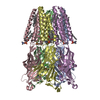
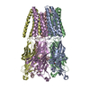
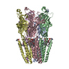
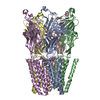
- Structure visualization
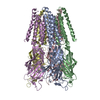
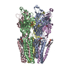
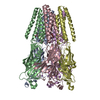
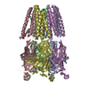
| Structure viewer | Molecule: MolmilJmol/JSmol |
|---|
- Downloads & links
-Download
| PDBx/mmCIF format | 6hya.cif.gz | 639.1 KB | Display | PDBx/mmCIF format |
|---|---|---|---|---|
| PDB format | pdb6hya.ent.gz | 538.5 KB | Display | PDB format |
| PDBx/mmJSON format | 6hya.json.gz | Tree view | PDBx/mmJSON format | |
| Others |  Other downloads Other downloads |
-Validation report
| Arichive directory | https://data.pdbj.org/pub/pdb/validation_reports/hy/6hyaftp://data.pdbj.org/pub/pdb/validation_reports/hy/6hya | HTTPS FTP |
|---|
-Related structure data
| Related structure data |  6hy5C  6hy9C  6hyrC  6hyvC  6hywC  6hyxC  6hyzC  6hz0C  6hz1C  6hz3C  6hzwC  6i08C C: citing same article ( |
|---|---|
| Similar structure data |


-Links
 PDBj
PDBj

- Assembly
| Deposited unit | 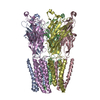
| ||||||||
|---|---|---|---|---|---|---|---|---|---|
| 1 |
| ||||||||
| Unit cell |
|
-Components
| #1: Protein | Mass: 36276.777 Da / Num. of mol.: 5 / Fragment: UNP RESIDUES 44-359 / Mutation: YES Source method: isolated from a genetically manipulated source Source: (gene. exp.) Gloeobacter violaceus PCC 7421 (bacteria)Gene: glvI, glr4197 / Production host: #2: Chemical | ChemComp-CL /   Mass: 35.453 Da / Num. of mol.: 5 / Source method: obtained synthetically / Formula: Cl Mass: 35.453 Da / Num. of mol.: 5 / Source method: obtained synthetically / Formula: Cl#3: Sugar | ChemComp-LMT / |   Type: D-saccharide / Mass: 510.615 Da / Num. of mol.: 1 / Source method: obtained synthetically / Formula: C24H46O11 / Comment: detergent*YM Type: D-saccharide / Mass: 510.615 Da / Num. of mol.: 1 / Source method: obtained synthetically / Formula: C24H46O11 / Comment: detergent*YM#4: Water | ChemComp-HOH / |  Mass: 18.015 Da / Num. of mol.: 1 / Source method: isolated from a natural source / Formula: H2O Mass: 18.015 Da / Num. of mol.: 1 / Source method: isolated from a natural source / Formula: H2O |
|---|
-Experimental details
-Experiment
| Experiment | Method: X-RAY DIFFRACTION / Number of used crystals: 1 |
|---|
- Sample preparation
| Crystal | Density Matthews: 4.99 Å3/Da / Density % sol: 75.36 % |
|---|---|
| Crystal grow | Temperature: 293 K / Method: evaporation / pH: 4 Details: PH 4, VAPOR DIFFUSION, TEMPERATURE 293K;12-14.5% PEG4K; 15% GLycerol; 400 mM NaSCN; 3% DMSO; 100mM NaAcetate pH 4. PH range: 4 |
-Data collection
| Diffraction | Mean temperature: 193 K / Serial crystal experiment: N |
|---|---|
| Diffraction source | Source: SYNCHROTRON / Site: SOLEIL  / Beamline: PROXIMA 1 / Wavelength: 0.9789 Å / Beamline: PROXIMA 1 / Wavelength: 0.9789 Å |
| Detector | Type: ADSC QUANTUM 315r / Detector: CCD / Date: Oct 11, 2015 |
| Radiation | Monochromator: CHANNEL CUT / Protocol: SINGLE WAVELENGTH / Monochromatic (M) / Laue (L): M / Scattering type: x-ray |
| Radiation wavelength | Wavelength: 0.9789 Å / Relative weight: 1 |
| Reflection | Resolution: 3.39→48.84 Å / Num. obs: 50723 / % possible obs: 99.1 % / Redundancy: 3.2 % / Biso Wilson estimate: 106.02 Å2 / Rmerge(I) obs: 0.065 / Net I/σ(I): 4.9 |
| Reflection shell | Resolution: 3.39→3.5 Å / Redundancy: 3.2 % / Rmerge(I) obs: 0.406 / Mean I/σ(I) obs: 1.4 / % possible all: 92.6 |
- Processing
| Software |
| ||||||||||||||||||||||||||||||||||||||||||||||||||||||||||||||||||||||||||||||||||||||||||||||||||||||||||||||||||||||||||||||||||||||||||||||||||||||
|---|---|---|---|---|---|---|---|---|---|---|---|---|---|---|---|---|---|---|---|---|---|---|---|---|---|---|---|---|---|---|---|---|---|---|---|---|---|---|---|---|---|---|---|---|---|---|---|---|---|---|---|---|---|---|---|---|---|---|---|---|---|---|---|---|---|---|---|---|---|---|---|---|---|---|---|---|---|---|---|---|---|---|---|---|---|---|---|---|---|---|---|---|---|---|---|---|---|---|---|---|---|---|---|---|---|---|---|---|---|---|---|---|---|---|---|---|---|---|---|---|---|---|---|---|---|---|---|---|---|---|---|---|---|---|---|---|---|---|---|---|---|---|---|---|---|---|---|---|---|---|---|
| Refinement | Method to determine structure: MOLECULAR REPLACEMENT / Resolution: 3.39→20 Å / Cor.coef. Fo:Fc: 0.923 / Cor.coef. Fo:Fc free: 0.9011 / Cross valid method: THROUGHOUT / σ(F): 0 / SU Rfree Blow DPI: 0.606
| ||||||||||||||||||||||||||||||||||||||||||||||||||||||||||||||||||||||||||||||||||||||||||||||||||||||||||||||||||||||||||||||||||||||||||||||||||||||
| Displacement parameters | Biso mean: 110.86 Å2
| ||||||||||||||||||||||||||||||||||||||||||||||||||||||||||||||||||||||||||||||||||||||||||||||||||||||||||||||||||||||||||||||||||||||||||||||||||||||
| Refine analyze | Luzzati coordinate error obs: 0.448 Å | ||||||||||||||||||||||||||||||||||||||||||||||||||||||||||||||||||||||||||||||||||||||||||||||||||||||||||||||||||||||||||||||||||||||||||||||||||||||
| Refinement step | Cycle: LAST / Resolution: 3.39→20 Å
| ||||||||||||||||||||||||||||||||||||||||||||||||||||||||||||||||||||||||||||||||||||||||||||||||||||||||||||||||||||||||||||||||||||||||||||||||||||||
| Refine LS restraints |
| ||||||||||||||||||||||||||||||||||||||||||||||||||||||||||||||||||||||||||||||||||||||||||||||||||||||||||||||||||||||||||||||||||||||||||||||||||||||
| LS refinement shell | Resolution: 3.39→3.53 Å / Total num. of bins used: 13
| ||||||||||||||||||||||||||||||||||||||||||||||||||||||||||||||||||||||||||||||||||||||||||||||||||||||||||||||||||||||||||||||||||||||||||||||||||||||
| Refinement TLS params. | Method: refined / Refine-ID: X-RAY DIFFRACTION
| ||||||||||||||||||||||||||||||||||||||||||||||||||||||||||||||||||||||||||||||||||||||||||||||||||||||||||||||||||||||||||||||||||||||||||||||||||||||
| Refinement TLS group |
|
