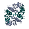
Evidence: scanning transmission electron microscopy, protein forms 2D lattices on bacterial surface and planar and tubular lattices in vitro., SAXS
Type
Name
Symmetry operation
Number
identity operation
1_555
x,y,z
1
Buried area
3230 Å2
ΔGint
-13 kcal/mol
Surface area
43050 Å2
Method
PISA
Unit cell
Length a, b, c (Å)
107.553, 115.114, 152.768
Angle α, β, γ (deg.)
90.00, 90.00, 90.00
Int Tables number
20
Space group name H-M
C2221
Components on special symmetry positions
ID
Model
Components
1
1
A-918-
HOH
-
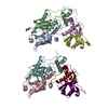
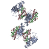
Components
#1: Protein
S-layerproteinsap / Surface array protein / Surface layer protein
Mass: 64944.977 Da / Num. of mol.: 1 Source method: isolated from a genetically manipulated source Source: (gene. exp.) Bacillus anthracis (anthrax bacterium) / Gene: sap, BA_0885, GBAA_0885, BAS0841 / Production host: Escherichia coli BL21 (bacteria) / References: UniProt: P49051
#2: Antibody
nanobodyAF684
Mass: 14110.615 Da / Num. of mol.: 1 Source method: isolated from a genetically manipulated source Source: (gene. exp.) Lama glama (llama) / Production host: Escherichia coli (E. coli) / Strain (production host): WK6
#3: Antibody
NanobodyAF694
Mass: 14913.369 Da / Num. of mol.: 1 Source method: isolated from a genetically manipulated source Source: (gene. exp.) Lama glama (llama) / Production host: Escherichia coli (E. coli) / Strain (production host): WK6
Resolution: 2.69→2.73 Å / Redundancy: 10.2 % / Mean I/σ(I) obs: 1.6 / Num. unique obs: 1282 / CC1/2: 0.658 / Rpim(I) all: 0.537 / % possible all: 96.8
-
Processing
Software
Name
Version
Classification
BUSTER
2.10.3
refinement
xia2
0.5.48
datareduction
xia2
0.5.48
datascaling
SHARP
2.8
phasing
Refinement
Method to determine structure: SAD / Resolution: 2.7→32.1 Å / Cor.coef. Fo:Fc: 0.946 / Cor.coef. Fo:Fc free: 0.914 / SU R Cruickshank DPI: 1.373 / Cross valid method: THROUGHOUT / σ(F): 0 / SU R Blow DPI: 1.305 / SU Rfree Blow DPI: 0.325 / SU Rfree Cruickshank DPI: 0.332
Rfactor
Num. reflection
% reflection
Selection details
Rfree
0.25
1304
4.95 %
RANDOM
Rwork
0.186
-
-
-
obs
0.19
26344
99.8 %
-
Displacement parameters
Biso mean: 65.23 Å2
Baniso -1
Baniso -2
Baniso -3
1-
-0.7087 Å2
0 Å2
0 Å2
2-
-
0.1676 Å2
0 Å2
3-
-
-
0.5411 Å2
Refine analyze
Luzzati coordinate error obs: 0.33 Å
Refinement step
Cycle: 1 / Resolution: 2.7→32.1 Å
Protein
Nucleic acid
Ligand
Solvent
Total
Num. atoms
6259
0
0
114
6373
Refine LS restraints
Refine-ID
Type
Dev ideal
Number
Restraint function
Weight
X-RAY DIFFRACTION
t_bond_d
0.01
6338
HARMONIC
2
X-RAY DIFFRACTION
t_angle_deg
1.38
8575
HARMONIC
2
X-RAY DIFFRACTION
t_dihedral_angle_d
2235
SINUSOIDAL
2
X-RAY DIFFRACTION
t_incorr_chiral_ct
X-RAY DIFFRACTION
t_pseud_angle
X-RAY DIFFRACTION
t_trig_c_planes
X-RAY DIFFRACTION
t_gen_planes
1071
HARMONIC
5
X-RAY DIFFRACTION
t_it
6338
HARMONIC
20
X-RAY DIFFRACTION
t_nbd
X-RAY DIFFRACTION
t_omega_torsion
3.16
X-RAY DIFFRACTION
t_other_torsion
23.19
X-RAY DIFFRACTION
t_improper_torsion
X-RAY DIFFRACTION
t_chiral_improper_torsion
861
SEMIHARMONIC
5
X-RAY DIFFRACTION
t_sum_occupancies
X-RAY DIFFRACTION
t_utility_distance
X-RAY DIFFRACTION
t_utility_angle
X-RAY DIFFRACTION
t_utility_torsion
X-RAY DIFFRACTION
t_ideal_dist_contact
6702
SEMIHARMONIC
4
LS refinement shell
Resolution: 2.7→2.81 Å / Total num. of bins used: 13
Rfactor
Num. reflection
% reflection
Rfree
0.3568
142
4.91 %
Rwork
0.2469
2750
-
all
0.2524
2892
-
obs
-
-
98.44 %
Refinement TLS params.
Method: refined / Refine-ID: X-RAY DIFFRACTION
ID
L11 (°2)
L12 (°2)
L13 (°2)
L22 (°2)
L23 (°2)
L33 (°2)
S11 (Å °)
S12 (Å °)
S13 (Å °)
S21 (Å °)
S22 (Å °)
S23 (Å °)
S31 (Å °)
S32 (Å °)
S33 (Å °)
T11 (Å2)
T12 (Å2)
T13 (Å2)
T22 (Å2)
T23 (Å2)
T33 (Å2)
Origin x (Å)
Origin y (Å)
Origin z (Å)
1
1.83
0.3491
-1.4318
0.7728
0.3164
3.9872
0.1618
0.0771
-0.1193
-0.1191
-0.0347
0.0765
-0.0371
0.1669
-0.127
-0.0139
-0.0116
0.061
-0.1226
0.0375
0.0832
152.944
96.8707
154.662
2
3.7805
1.159
-2.409
4.9206
-3.1652
3.9317
-0.034
0.0247
-0.0685
0.0266
0.052
0.1467
-0.1407
-0.0951
-0.018
-0.2232
-0.01
0.0194
-0.1602
-0.0523
0.1116
132.674
94.634
176.151
3
3.3626
-0.7058
-0.3775
4.7264
-1.5966
2.5104
0.1775
-0.219
0.7063
0.0739
-0.0211
-0.0002
-0.0793
-0.1333
-0.1564
-0.1667
0.0521
0.1458
-0.2336
-0.0656
0.1935
154.519
62.3985
174.975
4
1.673
0.6953
1.9859
1.7671
0.5858
7.1817
-0.2345
0.1095
-0.0957
-0.1361
0.2746
0.262
-0.3708
-0.1648
-0.0401
-0.192
0.021
-0.0706
-0.1798
0
0.1125
176.208
41.6472
145.351
5
1.004
0.1655
0.4203
1.3081
-0.5529
8.9093
0.2823
0.4911
-0.4883
-0.2485
0.2376
0.1586
0.9209
0.4699
-0.5199
-0.1583
0.1622
0.0367
-0.0274
-0.112
0.114
183.006
24.5225
146.497
6
0.8287
-1.3647
1.2383
0
0.492
5.3861
0.0931
0.1487
-0.524
0.12
-0.0049
0.4414
1.0885
1.0408
-0.0883
0.1929
0.2559
0.0285
-0.1815
-0.1344
0.087
185.224
13.8692
148.393
7
2.9668
-2.4947
-0.433
2.5917
1.118
1.7593
0.1905
-0.0659
0.4879
-0.0209
0.131
-0.2159
0.0942
-0.064
-0.3216
-0.2079
-0.0829
0.028
-0.2124
0.0234
0
162.227
30.2754
177.28
8
6.7387
-3.7576
-0.0257
3.269
-0.8756
2.3491
0.2408
-0.2365
0.1231
-0.2205
-0.1978
-0.2428
0.017
0.1955
-0.043
-0.1033
-0.0484
0.0332
-0.1415
0.0258
0.238
157.96
37.1058
179.963
9
3.7636
0.1735
1.9549
1.3787
-0.1326
3.2457
-0.1908
0.0635
0.0538
-0.0634
-0.0533
-0.2064
0.1884
0.3442
0.2442
-0.1236
-0.0061
0.057
-0.1791
0.0193
0.1385
160.815
97.465
178.939
10
0.8972
-0.8896
-0.3883
4.38
0.2807
2.1934
0.0898
0.2969
0.2392
0.1534
-0.1443
0.1243
-0.1114
-0.1305
0.0545
-0.088
0.0743
0.0012
-0.0958
0.0975
0.0279
155.721
108.017
131.728
Refinement TLS group
ID
Refine-ID
Refine TLS-ID
Selection details
1
X-RAY DIFFRACTION
1
{ A|216 - A|290 }
2
X-RAY DIFFRACTION
2
{ A|291 - A|386 }
3
X-RAY DIFFRACTION
3
{ A|387 - A|490 }
4
X-RAY DIFFRACTION
4
{ A|491 - A|592 }
5
X-RAY DIFFRACTION
5
{ A|593 - A|631 }
6
X-RAY DIFFRACTION
6
{ A|632 - A|696 }
7
X-RAY DIFFRACTION
7
{ A|697 - A|764 }
8
X-RAY DIFFRACTION
8
{ A|765 - A|811 }
9
X-RAY DIFFRACTION
9
{ G|1 - G|119 }
10
X-RAY DIFFRACTION
10
{ H|1 - H|123 }
+
About Yorodumi
-
News
-
Feb 9, 2022. New format data for meta-information of EMDB entries
New format data for meta-information of EMDB entries
Version 3 of the EMDB header file is now the official format.
The previous official version 1.9 will be removed from the archive.
In the structure databanks used in Yorodumi, some data are registered as the other names, "COVID-19 virus" and "2019-nCoV". Here are the details of the virus and the list of structure data.
Jan 31, 2019. EMDB accession codes are about to change! (news from PDBe EMDB page)
EMDB accession codes are about to change! (news from PDBe EMDB page)
The allocation of 4 digits for EMDB accession codes will soon come to an end. Whilst these codes will remain in use, new EMDB accession codes will include an additional digit and will expand incrementally as the available range of codes is exhausted. The current 4-digit format prefixed with “EMD-” (i.e. EMD-XXXX) will advance to a 5-digit format (i.e. EMD-XXXXX), and so on. It is currently estimated that the 4-digit codes will be depleted around Spring 2019, at which point the 5-digit format will come into force.
The EM Navigator/Yorodumi systems omit the EMD- prefix.
Related info.:Q: What is EMD? / ID/Accession-code notation in Yorodumi/EM Navigator
Yorodumi is a browser for structure data from EMDB, PDB, SASBDB, etc.
This page is also the successor to EM Navigator detail page, and also detail information page/front-end page for Omokage search.
The word "yorodu" (or yorozu) is an old Japanese word meaning "ten thousand". "mi" (miru) is to see.
Related info.:EMDB / PDB / SASBDB / Comparison of 3 databanks / Yorodumi Search / Aug 31, 2016. New EM Navigator & Yorodumi / Yorodumi Papers / Jmol/JSmol / Function and homology information / Changes in new EM Navigator and Yorodumi
 Movie
Movie Controller


 Open data
 Basic information
 Components
 Keywords
 Function and homology information

 X-RAY DIFFRACTION /
  Authors
 Belgium, 1items
  Citation
 Structure visualization
 Downloads & links
 Other downloads


 PDBj
PDBj

 Assembly
 Mass: 18.015 Da / Num. of mol.: 114 / Source method: isolated from a natural source / Formula: H2O
 Sample preparation
 / Beamline: I03 / Wavelength: 0.9795 Å
 Processing