 Movie
Movie Controller

[English] 日本語
 Yorodumi
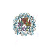
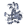
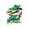
- PDB-2hue: Structure of the H3-H4 chaperone Asf1 bound to histones H3 and H4 -
+ Open data

- Basic information
| Entry | Database: PDB / ID: 2hue | ||||||
|---|---|---|---|---|---|---|---|
| Title | Structure of the H3-H4 chaperone Asf1 bound to histones H3 and H4 | ||||||
 Components Components |
| ||||||
 Keywords Keywords | DNA BINDING PROTEIN / mini beta sheet / elongated Beta sandwhich | ||||||
| Function / homology |  Function and homology information Function and homology informationFormation of Senescence-Associated Heterochromatin Foci (SAHF) / H3 histone acetyltransferase complex / acetyltransferase activator activity / DNA replication-dependent chromatin assembly / silent mating-type cassette heterochromatin formation / nucleosome disassembly / cellular response to stress / regulation of protein phosphorylation / negative regulation of DNA damage checkpoint / subtelomeric heterochromatin formation ...Formation of Senescence-Associated Heterochromatin Foci (SAHF) / H3 histone acetyltransferase complex / acetyltransferase activator activity / DNA replication-dependent chromatin assembly / silent mating-type cassette heterochromatin formation / nucleosome disassembly / cellular response to stress / regulation of protein phosphorylation / negative regulation of DNA damage checkpoint / subtelomeric heterochromatin formation / regulation of DNA repair / nucleosomal DNA binding / positive regulation of transcription elongation by RNA polymerase II / structural constituent of chromatin / nucleosome / nucleosome assembly / regulation of gene expression / chromatin organization / heterochromatin formation / histone binding / chromosome, telomeric region / chromatin remodeling / protein heterodimerization activity / regulation of transcription by RNA polymerase II / chromatin / DNA-templated transcription / DNA binding / nucleoplasm / nucleus / cytosol Similarity search - Function | ||||||
| Biological species |  | ||||||
| Method |  X-RAY DIFFRACTION / SYNCHROTRON / MOLECULAR REPLACEMENT / Resolution: 1.7 Å X-RAY DIFFRACTION / SYNCHROTRON / MOLECULAR REPLACEMENT / Resolution: 1.7 Å | ||||||
 Authors Authors | English, C.M. / Churchill, M.E.A. / Tyler, J.K. | ||||||
 Citation Citation | Journal: Cell(Cambridge,Mass.) / Year: 2006 Title: Structural basis for the histone chaperone activity of asf1. Authors: English, C.M. / Adkins, M.W. / Carson, J.J. / Churchill, M.E. / Tyler, J.K. | ||||||
| History |
| ||||||
| Remark 999 | SEQUENCE An Ala at position 102 agrees with the database reference GB P84233 |
- Structure visualization
| Structure viewer | Molecule: MolmilJmol/JSmol |
|---|
- Downloads & links
-Download
| PDBx/mmCIF format | 2hue.cif.gz | 90.6 KB | Display | PDBx/mmCIF format |
|---|---|---|---|---|
| PDB format | pdb2hue.ent.gz | 67.7 KB | Display | PDB format |
| PDBx/mmJSON format | 2hue.json.gz | Tree view | PDBx/mmJSON format | |
| Others |  Other downloads Other downloads |
-Validation report
| Arichive directory | https://data.pdbj.org/pub/pdb/validation_reports/hu/2hueftp://data.pdbj.org/pub/pdb/validation_reports/hu/2hue | HTTPS FTP |
|---|
-Related structure data
| Related structure data | |
|---|---|
| Similar structure data |


-Links
 PDBj
PDBj

- Assembly
| Deposited unit | 
| ||||||||
|---|---|---|---|---|---|---|---|---|---|
| 1 |
| ||||||||
| 2 | 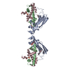
| ||||||||
| 3 | 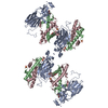
| ||||||||
| 4 | 
| ||||||||
| Unit cell |
| ||||||||
| Components on special symmetry positions |
|
-Components
-Protein , 3 types, 3 molecules ABC
| #1: Protein | Mass: 19663.973 Da / Num. of mol.: 1 / Fragment: residues 2-169 Source method: isolated from a genetically manipulated source Source: (gene. exp.) Gene: ASF1 / Plasmid: PST39 / Production host:  |
|---|---|
| #2: Protein | Mass: 8888.460 Da / Num. of mol.: 1 / Fragment: residues 62-136 / Mutation: G103A Source method: isolated from a genetically manipulated source Source: (gene. exp.) |
| #3: Protein | Mass: 9540.251 Da / Num. of mol.: 1 / Fragment: residues 20-102 Source method: isolated from a genetically manipulated source Source: (gene. exp.) |
-Non-polymers , 4 types, 372 molecules 


| #4: Chemical | ChemComp-ZN /  Mass: 65.409 Da / Num. of mol.: 1 / Source method: obtained synthetically / Formula: Zn Mass: 65.409 Da / Num. of mol.: 1 / Source method: obtained synthetically / Formula: Zn | ||||
|---|---|---|---|---|---|
| #5: Chemical |  Mass: 92.094 Da / Num. of mol.: 2 / Source method: obtained synthetically / Formula: C3H8O3 Mass: 92.094 Da / Num. of mol.: 2 / Source method: obtained synthetically / Formula: C3H8O3#6: Chemical |  Mass: 96.063 Da / Num. of mol.: 2 / Source method: obtained synthetically / Formula: SO4 Mass: 96.063 Da / Num. of mol.: 2 / Source method: obtained synthetically / Formula: SO4#7: Water | ChemComp-HOH / | Mass: 18.015 Da / Num. of mol.: 367 / Source method: isolated from a natural source / Formula: H2O |
-Experimental details
-Experiment
| Experiment | Method: X-RAY DIFFRACTION / Number of used crystals: 1 |
|---|
- Sample preparation
| Crystal | Density Matthews: 3.9 Å3/Da / Density % sol: 68.44 % |
|---|---|
| Crystal grow | Temperature: 295 K / Method: vapor diffusion, hanging drop / pH: 8.5 Details: 0.1M potassium sulfate, 0.1M Tris-HCl, 14.5% PEG 4K, pH 8.5, VAPOR DIFFUSION, HANGING DROP, temperature 295K |
-Data collection
| Diffraction | Mean temperature: 295 K | |||||||||
|---|---|---|---|---|---|---|---|---|---|---|
| Diffraction source | Source: SYNCHROTRON / Site: ALS  / Beamline: 4.2.2 / Wavelength: 1.000, 1.23 / Beamline: 4.2.2 / Wavelength: 1.000, 1.23 | |||||||||
| Detector | Type: NOIR-1 / Detector: CCD / Date: Sep 30, 2005 | |||||||||
| Radiation | Monochromator: Double crystal / Protocol: SINGLE WAVELENGTH / Monochromatic (M) / Laue (L): M / Scattering type: x-ray | |||||||||
| Radiation wavelength |
| |||||||||
| Reflection | Resolution: 1.7→12 Å / Num. obs: 61200 |
- Processing
| Software |
| ||||||||||||||||||
|---|---|---|---|---|---|---|---|---|---|---|---|---|---|---|---|---|---|---|---|
| Refinement | Method to determine structure: MOLECULAR REPLACEMENT / Resolution: 1.7→11.98 Å /
| ||||||||||||||||||
| Refinement step | Cycle: LAST / Resolution: 1.7→11.98 Å
| ||||||||||||||||||
| Refine LS restraints |
|
