 Movie
Movie Controller

[English] 日本語
 Yorodumi
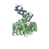
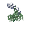
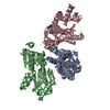
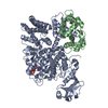
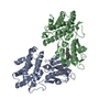
- PDB-6les: 3D domain-swapped dimer of the maltose-binding protein fused to a... -
+ Open data

- Basic information
| Entry | Database: PDB / ID: 6les | ||||||
|---|---|---|---|---|---|---|---|
| Title | 3D domain-swapped dimer of the maltose-binding protein fused to a fragment of the focal adhesion kinase | ||||||
 Components Components | Maltose/maltodextrin-binding periplasmic protein,Focal adhesion kinase 1 | ||||||
 Keywords Keywords | SUGAR BINDING PROTEIN / maltose binding protein / domain-swapping / arm exchange / folding / passenger protein / surface entropy reduction / fixed-arm carrier / dimer / FAK / apo-protein | ||||||
| Function / homology |  Function and homology information Function and homology informationnetrin-activated signaling pathway / regulation of substrate adhesion-dependent cell spreading / regulation of endothelial cell migration / regulation of epithelial cell migration / detection of muscle stretch / positive regulation of cell-cell adhesion mediated by integrin / JUN kinase binding / signal complex assembly / negative regulation of cell adhesion mediated by integrin / positive regulation of macrophage proliferation ...netrin-activated signaling pathway / regulation of substrate adhesion-dependent cell spreading / regulation of endothelial cell migration / regulation of epithelial cell migration / detection of muscle stretch / positive regulation of cell-cell adhesion mediated by integrin / JUN kinase binding / signal complex assembly / negative regulation of cell adhesion mediated by integrin / positive regulation of macrophage proliferation / DCC mediated attractive signaling / positive regulation of fibroblast migration / negative regulation of cell-cell adhesion / Signal regulatory protein family interactions / MET activates PTK2 signaling / growth hormone receptor signaling pathway / positive regulation of ubiquitin-dependent protein catabolic process / regulation of focal adhesion assembly / Apoptotic cleavage of cellular proteins / p130Cas linkage to MAPK signaling for integrins / positive regulation of wound healing / regulation of osteoblast differentiation / establishment of cell polarity / Fc-gamma receptor signaling pathway involved in phagocytosis / regulation of cytoskeleton organization / positive regulation of macrophage chemotaxis / vascular endothelial cell response to oscillatory fluid shear stress / detection of maltose stimulus / GRB2:SOS provides linkage to MAPK signaling for Integrins / maltose transport complex / carbohydrate transport / positive regulation of epithelial cell migration / negative regulation of anoikis / ephrin receptor signaling pathway / vascular endothelial growth factor receptor signaling pathway / Estrogen-dependent nuclear events downstream of ESR-membrane signaling / heart morphogenesis / carbohydrate transmembrane transporter activity / regulation of cell adhesion / maltose binding / RHO GTPases Activate WASPs and WAVEs / maltose transport / maltodextrin transmembrane transport / positive regulation of epithelial to mesenchymal transition / ATP-binding cassette (ABC) transporter complex, substrate-binding subunit-containing / Integrin signaling / stress fiber / transforming growth factor beta receptor signaling pathway / EPHB-mediated forward signaling / placenta development / ATP-binding cassette (ABC) transporter complex / protein tyrosine phosphatase activity / NCAM signaling for neurite out-growth / SH2 domain binding / axon guidance / Turbulent (oscillatory, disturbed) flow shear stress activates signaling by PIEZO1 and integrins in endothelial cells / integrin-mediated signaling pathway / molecular function activator activity / cell motility / non-specific protein-tyrosine kinase / FCGR3A-mediated phagocytosis / cell chemotaxis / non-membrane spanning protein tyrosine kinase activity / Regulation of actin dynamics for phagocytic cup formation / VEGFA-VEGFR2 Pathway / integrin binding / epidermal growth factor receptor signaling pathway / regulation of cell shape / regulation of cell population proliferation / cell migration / outer membrane-bounded periplasmic space / RAF/MAP kinase cascade / actin binding / protein tyrosine kinase activity / angiogenesis / protein phosphatase binding / cell cortex / cytoskeleton / ciliary basal body / positive regulation of phosphatidylinositol 3-kinase/protein kinase B signal transduction / periplasmic space / Extra-nuclear estrogen signaling / positive regulation of cell migration / focal adhesion / centrosome / positive regulation of cell population proliferation / DNA damage response / negative regulation of apoptotic process / protein kinase binding / perinuclear region of cytoplasm / ATP binding / membrane / nucleus / plasma membrane / cytosol Similarity search - Function | ||||||
| Biological species |   Homo sapiens (human) Homo sapiens (human) | ||||||
| Method |  X-RAY DIFFRACTION / SYNCHROTRON / MOLECULAR REPLACEMENT / Resolution: 2.004 Å X-RAY DIFFRACTION / SYNCHROTRON / MOLECULAR REPLACEMENT / Resolution: 2.004 Å | ||||||
 Authors Authors | Momin, A.A. / Shahul Hameed, U.F. / Arold, S.T. | ||||||
| Funding support |  Saudi Arabia, 1items Saudi Arabia, 1items
| ||||||
 Citation Citation | Journal: Sci Rep / Year: 2019 Title: Passenger sequences can promote interlaced dimers in a common variant of the maltose-binding protein. Authors: Momin, A.A. / Hameed, U.F.S. / Arold, S.T. | ||||||
| History |
|
- Structure visualization
| Structure viewer | Molecule: MolmilJmol/JSmol |
|---|
- Downloads & links
-Download
| PDBx/mmCIF format | 6les.cif.gz | 575 KB | Display | PDBx/mmCIF format |
|---|---|---|---|---|
| PDB format | pdb6les.ent.gz | 476.5 KB | Display | PDB format |
| PDBx/mmJSON format | 6les.json.gz | Tree view | PDBx/mmJSON format | |
| Others |  Other downloads Other downloads |
-Validation report
| Arichive directory | https://data.pdbj.org/pub/pdb/validation_reports/le/6lesftp://data.pdbj.org/pub/pdb/validation_reports/le/6les | HTTPS FTP |
|---|
-Related structure data
| Related structure data | 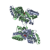 6lf3C 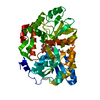 5bmyS C: citing same article ( S: Starting model for refinement |
|---|---|
| Similar structure data |


-Links
 PDBj
PDBj


- Assembly
| Deposited unit | 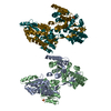
| ||||||||
|---|---|---|---|---|---|---|---|---|---|
| 1 | 
| ||||||||
| 2 | 
| ||||||||
| Unit cell |
|
-Components
| #1: Protein | Mass: 43853.605 Da / Num. of mol.: 4 Mutation: surface entropy reduction mutant,D83A,K84A,E173A,N174A,K240A,E360A,K363A,D364A Source method: isolated from a genetically manipulated source Details: residues 805-832 of the human focal adhesion kinase are C-terminally fused to the maltose-binding protein Source: (gene. exp.) Homo sapiens (human)Gene: malE, b4034, JW3994, PTK2, FAK, FAK1 / Plasmid: pJEx411c / Production host: References: UniProt: P0AEX9, UniProt: Q05397, non-specific protein-tyrosine kinase #2: Chemical |   Mass: 96.063 Da / Num. of mol.: 2 / Source method: obtained synthetically / Formula: SO4 Mass: 96.063 Da / Num. of mol.: 2 / Source method: obtained synthetically / Formula: SO4#3: Water | ChemComp-HOH / |  Mass: 18.015 Da / Num. of mol.: 428 / Source method: isolated from a natural source / Formula: H2O Mass: 18.015 Da / Num. of mol.: 428 / Source method: isolated from a natural source / Formula: H2OHas ligand of interest | N | |
|---|
-Experimental details
-Experiment
| Experiment | Method: X-RAY DIFFRACTION / Number of used crystals: 1 |
|---|
- Sample preparation
| Crystal | Density Matthews: 2.35 Å3/Da / Density % sol: 42.5 % |
|---|---|
| Crystal grow | Temperature: 298 K / Method: vapor diffusion, hanging drop / pH: 10.5 Details: 2000mM Ammonium sulfate, 100mM CAPS/ Sodium hydroxide pH 10.5 |
-Data collection
| Diffraction | Mean temperature: 100 K / Serial crystal experiment: N |
|---|---|
| Diffraction source | Source: SYNCHROTRON / Site: SOLEIL  / Beamline: PROXIMA 2 / Wavelength: 0.98 Å / Beamline: PROXIMA 2 / Wavelength: 0.98 Å |
| Detector | Type: DECTRIS EIGER X 9M / Detector: PIXEL / Date: Jul 13, 2019 |
| Radiation | Protocol: SINGLE WAVELENGTH / Monochromatic (M) / Laue (L): M / Scattering type: x-ray |
| Radiation wavelength | Wavelength: 0.98 Å / Relative weight: 1 |
| Reflection | Resolution: 2→47.49 Å / Num. obs: 93102 / % possible obs: 95.33 % / Redundancy: 8.12 % / Biso Wilson estimate: 46.1 Å2 / CC1/2: 0.995 / Rmerge(I) obs: 0.1033 / Rpim(I) all: 0.06574 / Rrim(I) all: 0.1229 / Χ2: 0.86 / Net I/σ(I): 8.07 |
| Reflection shell | Resolution: 2.004→2.076 Å / Redundancy: 1.2 % / Rmerge(I) obs: 0.944 / Mean I/σ(I) obs: 1.2 / Num. unique obs: 8178 / CC1/2: 0.6 / Rpim(I) all: 0.7233 / Χ2: 0.55 / % possible all: 84.49 |
- Processing
| Software |
| |||||||||||||||||||||||||||||||||||||||||||||||||||||||||||||||||||||||||||||||||||||||||||||||||||||||||||||||||||||||||||||||||||||||||||||||||||||||||||||||||||||||||||||||||||||||||||||||||||||||||||||||||||||||||||||||||||||||||||||||||||||||||||||||||||||||||||||||||||||||||||||||||||||||||||||||||||||||||||||||||||||||||||||||||||||||||||||||||||||||||||||||||||||||||||||||||||||||||||||||||||||||||||||||||||||||||||||||||||||||||||||||||||||||||||||||||||||||||||||||||||||||||||||||||||||||||||||||||||||||||||||
|---|---|---|---|---|---|---|---|---|---|---|---|---|---|---|---|---|---|---|---|---|---|---|---|---|---|---|---|---|---|---|---|---|---|---|---|---|---|---|---|---|---|---|---|---|---|---|---|---|---|---|---|---|---|---|---|---|---|---|---|---|---|---|---|---|---|---|---|---|---|---|---|---|---|---|---|---|---|---|---|---|---|---|---|---|---|---|---|---|---|---|---|---|---|---|---|---|---|---|---|---|---|---|---|---|---|---|---|---|---|---|---|---|---|---|---|---|---|---|---|---|---|---|---|---|---|---|---|---|---|---|---|---|---|---|---|---|---|---|---|---|---|---|---|---|---|---|---|---|---|---|---|---|---|---|---|---|---|---|---|---|---|---|---|---|---|---|---|---|---|---|---|---|---|---|---|---|---|---|---|---|---|---|---|---|---|---|---|---|---|---|---|---|---|---|---|---|---|---|---|---|---|---|---|---|---|---|---|---|---|---|---|---|---|---|---|---|---|---|---|---|---|---|---|---|---|---|---|---|---|---|---|---|---|---|---|---|---|---|---|---|---|---|---|---|---|---|---|---|---|---|---|---|---|---|---|---|---|---|---|---|---|---|---|---|---|---|---|---|---|---|---|---|---|---|---|---|---|---|---|---|---|---|---|---|---|---|---|---|---|---|---|---|---|---|---|---|---|---|---|---|---|---|---|---|---|---|---|---|---|---|---|---|---|---|---|---|---|---|---|---|---|---|---|---|---|---|---|---|---|---|---|---|---|---|---|---|---|---|---|---|---|---|---|---|---|---|---|---|---|---|---|---|---|---|---|---|---|---|---|---|---|---|---|---|---|---|---|---|---|---|---|---|---|---|---|---|---|---|---|---|---|---|---|---|---|---|---|---|---|---|---|---|---|---|---|---|---|---|---|---|---|---|---|---|---|---|---|---|---|---|---|---|---|---|---|---|---|---|---|---|---|---|---|---|---|---|---|---|---|---|---|---|---|---|---|---|---|---|---|---|---|---|---|---|---|---|---|---|---|---|---|---|---|---|---|---|---|---|---|---|---|---|---|---|---|---|---|---|---|---|---|---|---|---|---|---|---|---|---|---|---|---|---|---|---|---|---|---|---|---|---|---|---|---|---|---|---|---|---|---|---|---|---|---|---|---|---|---|---|---|---|---|---|---|---|---|---|---|---|---|---|---|---|---|---|---|
| Refinement | Method to determine structure: MOLECULAR REPLACEMENT Starting model: 5BMY Resolution: 2.004→47.45 Å / Cor.coef. Fo:Fc: 0.964 / Cor.coef. Fo:Fc free: 0.951 / SU B: 13.811 / SU ML: 0.175 / Cross valid method: THROUGHOUT / ESU R: 0.221 / ESU R Free: 0.174 Details: U VALUES : WITH TLS ADDED HYDROGENS HAVE BEEN ADDED IN THE RIDING POSITIONS U VALUES : RESIDUAL ONLY
| |||||||||||||||||||||||||||||||||||||||||||||||||||||||||||||||||||||||||||||||||||||||||||||||||||||||||||||||||||||||||||||||||||||||||||||||||||||||||||||||||||||||||||||||||||||||||||||||||||||||||||||||||||||||||||||||||||||||||||||||||||||||||||||||||||||||||||||||||||||||||||||||||||||||||||||||||||||||||||||||||||||||||||||||||||||||||||||||||||||||||||||||||||||||||||||||||||||||||||||||||||||||||||||||||||||||||||||||||||||||||||||||||||||||||||||||||||||||||||||||||||||||||||||||||||||||||||||||||||||||||||||
| Solvent computation | Ion probe radii: 0.7 Å / Shrinkage radii: 0.7 Å / VDW probe radii: 1.1 Å | |||||||||||||||||||||||||||||||||||||||||||||||||||||||||||||||||||||||||||||||||||||||||||||||||||||||||||||||||||||||||||||||||||||||||||||||||||||||||||||||||||||||||||||||||||||||||||||||||||||||||||||||||||||||||||||||||||||||||||||||||||||||||||||||||||||||||||||||||||||||||||||||||||||||||||||||||||||||||||||||||||||||||||||||||||||||||||||||||||||||||||||||||||||||||||||||||||||||||||||||||||||||||||||||||||||||||||||||||||||||||||||||||||||||||||||||||||||||||||||||||||||||||||||||||||||||||||||||||||||||||||||
| Displacement parameters | Biso max: 129.9 Å2 / Biso mean: 50.329 Å2 / Biso min: 26.93 Å2
| |||||||||||||||||||||||||||||||||||||||||||||||||||||||||||||||||||||||||||||||||||||||||||||||||||||||||||||||||||||||||||||||||||||||||||||||||||||||||||||||||||||||||||||||||||||||||||||||||||||||||||||||||||||||||||||||||||||||||||||||||||||||||||||||||||||||||||||||||||||||||||||||||||||||||||||||||||||||||||||||||||||||||||||||||||||||||||||||||||||||||||||||||||||||||||||||||||||||||||||||||||||||||||||||||||||||||||||||||||||||||||||||||||||||||||||||||||||||||||||||||||||||||||||||||||||||||||||||||||||||||||||
| Refinement step | Cycle: final / Resolution: 2.004→47.45 Å
| |||||||||||||||||||||||||||||||||||||||||||||||||||||||||||||||||||||||||||||||||||||||||||||||||||||||||||||||||||||||||||||||||||||||||||||||||||||||||||||||||||||||||||||||||||||||||||||||||||||||||||||||||||||||||||||||||||||||||||||||||||||||||||||||||||||||||||||||||||||||||||||||||||||||||||||||||||||||||||||||||||||||||||||||||||||||||||||||||||||||||||||||||||||||||||||||||||||||||||||||||||||||||||||||||||||||||||||||||||||||||||||||||||||||||||||||||||||||||||||||||||||||||||||||||||||||||||||||||||||||||||||
| LS refinement shell | Resolution: 2.004→2.056 Å / Rfactor Rfree error: 0
| |||||||||||||||||||||||||||||||||||||||||||||||||||||||||||||||||||||||||||||||||||||||||||||||||||||||||||||||||||||||||||||||||||||||||||||||||||||||||||||||||||||||||||||||||||||||||||||||||||||||||||||||||||||||||||||||||||||||||||||||||||||||||||||||||||||||||||||||||||||||||||||||||||||||||||||||||||||||||||||||||||||||||||||||||||||||||||||||||||||||||||||||||||||||||||||||||||||||||||||||||||||||||||||||||||||||||||||||||||||||||||||||||||||||||||||||||||||||||||||||||||||||||||||||||||||||||||||||||||||||||||||
| Refinement TLS params. | Method: refined / Refine-ID: X-RAY DIFFRACTION
| |||||||||||||||||||||||||||||||||||||||||||||||||||||||||||||||||||||||||||||||||||||||||||||||||||||||||||||||||||||||||||||||||||||||||||||||||||||||||||||||||||||||||||||||||||||||||||||||||||||||||||||||||||||||||||||||||||||||||||||||||||||||||||||||||||||||||||||||||||||||||||||||||||||||||||||||||||||||||||||||||||||||||||||||||||||||||||||||||||||||||||||||||||||||||||||||||||||||||||||||||||||||||||||||||||||||||||||||||||||||||||||||||||||||||||||||||||||||||||||||||||||||||||||||||||||||||||||||||||||||||||||
| Refinement TLS group |
|
