 Movie
Movie Controller


+ Open data

- Basic information
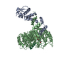
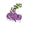
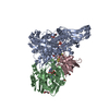
| Entry | Database: PDB / ID: 2p1q | ||||||
|---|---|---|---|---|---|---|---|
| Title | Mechanism of Auxin Perception by the TIR1 ubiquitin ligase | ||||||
 Components Components |
| ||||||
 Keywords Keywords | SIGNALING PROTEIN / F-box / leucine rich repeat | ||||||
| Function / homology |  Function and homology information Function and homology informationauxin receptor activity / pollen maturation / auxin binding / lateral root formation / gravitropism / stamen development / phragmoplast / ethylene-activated signaling pathway / jasmonic acid mediated signaling pathway / response to auxin ...auxin receptor activity / pollen maturation / auxin binding / lateral root formation / gravitropism / stamen development / phragmoplast / ethylene-activated signaling pathway / jasmonic acid mediated signaling pathway / response to auxin / response to jasmonic acid / male meiotic nuclear division / auxin-activated signaling pathway / negative regulation of DNA recombination / response to water deprivation / cellular response to phosphate starvation / inositol hexakisphosphate binding / SCF ubiquitin ligase complex / SCF-dependent proteasomal ubiquitin-dependent protein catabolic process / cullin family protein binding / ubiquitin ligase complex / chromosome segregation / defense response / response to wounding / microtubule cytoskeleton organization / spindle / ubiquitin-protein transferase activity / mitotic cell cycle / ubiquitin-dependent protein catabolic process / transcription cis-regulatory region binding / protein ubiquitination / DNA-binding transcription factor activity / mitochondrion / nucleus / cytoplasm / cytosol Similarity search - Function | ||||||
| Biological species |  | ||||||
| Method |  X-RAY DIFFRACTION / SYNCHROTRON / MOLECULAR REPLACEMENT / Resolution: 1.91 Å X-RAY DIFFRACTION / SYNCHROTRON / MOLECULAR REPLACEMENT / Resolution: 1.91 Å | ||||||
 Authors Authors | Tan, X. / Calderon-Villalobos, L.I.A. / Sharon, M. / Robinson, C.V. / Estelle, M. / Zheng, N. | ||||||
 Citation Citation | Journal: Nature / Year: 2007 Title: Mechanism of auxin perception by the TIR1 ubiquitin ligase. Authors: Tan, X. / Calderon-Villalobos, L.I. / Sharon, M. / Zheng, C. / Robinson, C.V. / Estelle, M. / Zheng, N. | ||||||
| History |
|
- Structure visualization
| Structure viewer | Molecule: MolmilJmol/JSmol |
|---|
- Downloads & links
-Download
| PDBx/mmCIF format | 2p1q.cif.gz | 172.5 KB | Display | PDBx/mmCIF format |
|---|---|---|---|---|
| PDB format | pdb2p1q.ent.gz | 131.9 KB | Display | PDB format |
| PDBx/mmJSON format | 2p1q.json.gz | Tree view | PDBx/mmJSON format | |
| Others |  Other downloads Other downloads |
-Validation report
| Arichive directory | https://data.pdbj.org/pub/pdb/validation_reports/p1/2p1qftp://data.pdbj.org/pub/pdb/validation_reports/p1/2p1q | HTTPS FTP |
|---|
-Related structure data
| Related structure data |  2p1mSC  2p1nC  2p1oC  2p1pC S: Starting model for refinement C: citing same article ( |
|---|---|
| Similar structure data |


-Links
 PDBj
PDBj


- Assembly
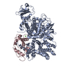
| Deposited unit | 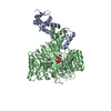
| ||||||||
|---|---|---|---|---|---|---|---|---|---|
| 1 |
| ||||||||
| 2 | 
| ||||||||
| Unit cell |
| ||||||||
| Components on special symmetry positions |
|
-Components
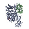
-Protein , 2 types, 2 molecules AB
| #1: Protein | Mass: 17876.043 Da / Num. of mol.: 1 Source method: isolated from a genetically manipulated source Source: (gene. exp.)   Spodoptera frugiperda (fall armyworm) / References: UniProt: Q39255 Spodoptera frugiperda (fall armyworm) / References: UniProt: Q39255 |
|---|---|
| #2: Protein | Mass: 66875.438 Da / Num. of mol.: 1 Source method: isolated from a genetically manipulated source Source: (gene. exp.) Spodoptera frugiperda (fall armyworm) / References: UniProt: Q570C0 |
-Protein/peptide , 1 types, 1 molecules C
| #3: Protein/peptide | Mass: 1601.872 Da / Num. of mol.: 1 / Source method: obtained synthetically Details: IAA7 peptide and indole-3-acetic acid are from commercial sources. It naturally occurs in Arabidopsis thaliana References: UniProt: Q38825 |
|---|
-Non-polymers , 3 types, 772 molecules 

| #4: Chemical | ChemComp-IHP /  Mass: 660.035 Da / Num. of mol.: 1 / Source method: obtained synthetically / Formula: C6H18O24P6 Mass: 660.035 Da / Num. of mol.: 1 / Source method: obtained synthetically / Formula: C6H18O24P6 |
|---|---|
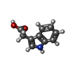
| #5: Chemical | ChemComp-IAC /  Mass: 175.184 Da / Num. of mol.: 1 / Source method: obtained synthetically / Formula: C10H9NO2 Mass: 175.184 Da / Num. of mol.: 1 / Source method: obtained synthetically / Formula: C10H9NO2 |
| #6: Water | ChemComp-HOH / Mass: 18.015 Da / Num. of mol.: 770 / Source method: isolated from a natural source / Formula: H2O |
-Experimental details
-Experiment
| Experiment | Method: X-RAY DIFFRACTION / Number of used crystals: 1 |
|---|
- Sample preparation
| Crystal | Density Matthews: 2.93 Å3/Da / Density % sol: 58.02 % |
|---|---|
| Crystal grow | Temperature: 298 K / Method: vapor diffusion, hanging drop / pH: 6 Details: 100 mM BTP, 10% 14% PEG 20,000, 200 mM NaCl, and 5 mM DTT, pH 6, VAPOR DIFFUSION, HANGING DROP, temperature 298K |
-Data collection
| Diffraction | Mean temperature: 100 K |
|---|---|
| Diffraction source | Source: SYNCHROTRON / Site: ALS  / Beamline: 8.2.1 / Wavelength: 1 Å / Beamline: 8.2.1 / Wavelength: 1 Å |
| Detector | Type: ADSC QUANTUM 315 / Detector: CCD / Date: Sep 1, 2006 |
| Radiation | Protocol: SINGLE WAVELENGTH / Monochromatic (M) / Laue (L): M / Scattering type: x-ray |
| Radiation wavelength | Wavelength: 1 Å / Relative weight: 1 |
| Reflection | Resolution: 1.91→50 Å / Num. all: 69793 / Num. obs: 69793 / % possible obs: 99.9 % / Redundancy: 3.8 % / Rsym value: 0.039 / Net I/σ(I): 33 |
| Reflection shell | Resolution: 1.91→2.03 Å / Redundancy: 3.6 % / Mean I/σ(I) obs: 4.9 / Rsym value: 0.249 / % possible all: 100 |
- Processing
| Software |
| ||||||||||||||||||||||||||||||||||||||||||||||||||||||||||||||||||||||||||||||||||||||||||||||||||||||||||||||||||||||||||||||||||||||||||||||||||||||||||||||||||||||||||
|---|---|---|---|---|---|---|---|---|---|---|---|---|---|---|---|---|---|---|---|---|---|---|---|---|---|---|---|---|---|---|---|---|---|---|---|---|---|---|---|---|---|---|---|---|---|---|---|---|---|---|---|---|---|---|---|---|---|---|---|---|---|---|---|---|---|---|---|---|---|---|---|---|---|---|---|---|---|---|---|---|---|---|---|---|---|---|---|---|---|---|---|---|---|---|---|---|---|---|---|---|---|---|---|---|---|---|---|---|---|---|---|---|---|---|---|---|---|---|---|---|---|---|---|---|---|---|---|---|---|---|---|---|---|---|---|---|---|---|---|---|---|---|---|---|---|---|---|---|---|---|---|---|---|---|---|---|---|---|---|---|---|---|---|---|---|---|---|---|---|---|---|
| Refinement | Method to determine structure: MOLECULAR REPLACEMENT Starting model: PDB ENTRY: 2P1M Resolution: 1.91→50 Å / Cor.coef. Fo:Fc: 0.963 / Cor.coef. Fo:Fc free: 0.946 / SU B: 5.47 / SU ML: 0.086 / Cross valid method: THROUGHOUT / ESU R: 0.129 / ESU R Free: 0.128 / Stereochemistry target values: MAXIMUM LIKELIHOOD / Details: HYDROGENS HAVE BEEN ADDED IN THE RIDING POSITIONS
| ||||||||||||||||||||||||||||||||||||||||||||||||||||||||||||||||||||||||||||||||||||||||||||||||||||||||||||||||||||||||||||||||||||||||||||||||||||||||||||||||||||||||||
| Solvent computation | Ion probe radii: 0.8 Å / Shrinkage radii: 0.8 Å / VDW probe radii: 1.2 Å / Solvent model: MASK | ||||||||||||||||||||||||||||||||||||||||||||||||||||||||||||||||||||||||||||||||||||||||||||||||||||||||||||||||||||||||||||||||||||||||||||||||||||||||||||||||||||||||||
| Displacement parameters | Biso mean: 27.399 Å2
| ||||||||||||||||||||||||||||||||||||||||||||||||||||||||||||||||||||||||||||||||||||||||||||||||||||||||||||||||||||||||||||||||||||||||||||||||||||||||||||||||||||||||||
| Refinement step | Cycle: LAST / Resolution: 1.91→50 Å
| ||||||||||||||||||||||||||||||||||||||||||||||||||||||||||||||||||||||||||||||||||||||||||||||||||||||||||||||||||||||||||||||||||||||||||||||||||||||||||||||||||||||||||
| Refine LS restraints |
| ||||||||||||||||||||||||||||||||||||||||||||||||||||||||||||||||||||||||||||||||||||||||||||||||||||||||||||||||||||||||||||||||||||||||||||||||||||||||||||||||||||||||||
| LS refinement shell | Resolution: 1.91→1.956 Å / Total num. of bins used: 20
|
