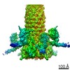
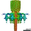
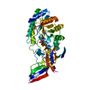
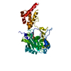
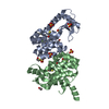
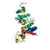
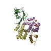
Journal: EMBO J / Year: 2019 Title: Structure and transformation of bacteriophage A511 baseplate and tail upon infection of cells. Authors: Ricardo C Guerrero-Ferreira / Mario Hupfeld / Sergey Nazarov / Nicholas Mi Taylor / Mikhail M Shneider / Jagan M Obbineni / Martin J Loessner / Takashi Ishikawa / Jochen Klumpp / Petr G Leiman / Abstract: Contractile injection systems (bacteriophage tails, type VI secretions system, R-type pyocins, etc.) utilize a rigid tube/contractile sheath assembly for breaching the envelope of bacterial and ...Contractile injection systems (bacteriophage tails, type VI secretions system, R-type pyocins, etc.) utilize a rigid tube/contractile sheath assembly for breaching the envelope of bacterial and eukaryotic cells. Among contractile injection systems, bacteriophages that infect Gram-positive bacteria represent the least understood members. Here, we describe the structure of bacteriophage A511 tail in its pre- and post-host attachment states (extended and contracted, respectively) using cryo-electron microscopy, cryo-electron tomography, and X-ray crystallography. We show that the structure of the tube-baseplate complex of A511 is similar to that of phage T4, but the A511 baseplate is decorated with different receptor-binding proteins, which undergo a large structural transformation upon host attachment and switch the symmetry of the baseplate-tail fiber assembly from threefold to sixfold. For the first time under native conditions, we show that contraction of the phage tail sheath assembly starts at the baseplate and propagates through the sheath in a domino-like motion.
In the structure databanks used in Yorodumi, some data are registered as the other names, "COVID-19 virus" and "2019-nCoV". Here are the details of the virus and the list of structure data.
Jan 31, 2019. EMDB accession codes are about to change! (news from PDBe EMDB page)
EMDB accession codes are about to change! (news from PDBe EMDB page)
The allocation of 4 digits for EMDB accession codes will soon come to an end. Whilst these codes will remain in use, new EMDB accession codes will include an additional digit and will expand incrementally as the available range of codes is exhausted. The current 4-digit format prefixed with “EMD-” (i.e. EMD-XXXX) will advance to a 5-digit format (i.e. EMD-XXXXX), and so on. It is currently estimated that the 4-digit codes will be depleted around Spring 2019, at which point the 5-digit format will come into force.
The EM Navigator/Yorodumi systems omit the EMD- prefix.
Related info.:Q: What is EMD? / ID/Accession-code notation in Yorodumi/EM Navigator
Yorodumi is a browser for structure data from EMDB, PDB, SASBDB, etc.
This page is also the successor to EM Navigator detail page, and also detail information page/front-end page for Omokage search.
The word "yorodu" (or yorozu) is an old Japanese word meaning "ten thousand". "mi" (miru) is to see.
Related info.:EMDB / PDB / SASBDB / Comparison of 3 databanks / Yorodumi Search / Aug 31, 2016. New EM Navigator & Yorodumi / Yorodumi Papers / Jmol/JSmol / Function and homology information / Changes in new EM Navigator and Yorodumi
 Movie
Movie Controller


 Open data
 Basic information
 Components
 Keywords
 Function and homology information
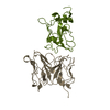
 Listeria phage A511 (virus)
 X-RAY DIFFRACTION /
  Authors
 Switzerland, 1items
  Citation

 Structure visualization
 Downloads & links
 Other downloads
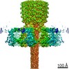


 PDBj
PDBj Assembly
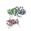


 Mass: 35.453 Da / Num. of mol.: 1 / Source method: obtained synthetically / Formula: Cl
 Mass: 18.015 Da / Num. of mol.: 95 / Source method: isolated from a natural source / Formula: H2O
 Sample preparation
 Processing