 Movie
Movie Controller

[English] 日本語
 Yorodumi
- PDB-3ctl: Crystal structure of D-Allulose 6-Phosphate 3-Epimerase from Esch... -
+ Open data

- Basic information
| Entry | Database: PDB / ID: 3ctl | ||||||
|---|---|---|---|---|---|---|---|
| Title | Crystal structure of D-Allulose 6-Phosphate 3-Epimerase from Escherichia coli K12 complexed with D-glucitol 6-phosphate and magnesium | ||||||
 Components Components | D-allulose-6-phosphate 3-epimerase | ||||||
 Keywords Keywords | ISOMERASE / D-Allulose 6-Phosphate 3-Epimerase / D-glucitol 6-phosphate / (beta/alpha)8 barrel / Carbohydrate metabolism | ||||||
| Function / homology |  Function and homology information Function and homology informationallulose 6-phosphate 3-epimerase activity / D-allose catabolic process / D-ribulose-phosphate 3-epimerase activity / Isomerases; Racemases and epimerases; Acting on carbohydrates and derivatives / pentose-phosphate shunt, non-oxidative branch / carbohydrate metabolic process / metal ion binding / cytosol Similarity search - Function | ||||||
| Biological species |  | ||||||
| Method |  X-RAY DIFFRACTION / SYNCHROTRON / MOLECULAR REPLACEMENT / Resolution: 2.2 Å X-RAY DIFFRACTION / SYNCHROTRON / MOLECULAR REPLACEMENT / Resolution: 2.2 Å | ||||||
 Authors Authors | Fedorov, A.A. / Fedorov, E.V. / Chan, K.K. / Gerlt, J.A. / Almo, S.C. | ||||||
 Citation Citation | Journal: Biochemistry / Year: 2008 Title: Structural basis for substrate specificity in phosphate binding (beta/alpha)8-barrels: D-allulose 6-phosphate 3-epimerase from Escherichia coli K-12. Authors: Chan, K.K. / Fedorov, A.A. / Fedorov, E.V. / Almo, S.C. / Gerlt, J.A. | ||||||
| History |
|
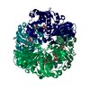
- Structure visualization
| Structure viewer | Molecule: MolmilJmol/JSmol |
|---|
- Downloads & links
-Download
| PDBx/mmCIF format | 3ctl.cif.gz | 266.9 KB | Display | PDBx/mmCIF format |
|---|---|---|---|---|
| PDB format | pdb3ctl.ent.gz | 218.9 KB | Display | PDB format |
| PDBx/mmJSON format | 3ctl.json.gz | Tree view | PDBx/mmJSON format | |
| Others |  Other downloads Other downloads |
-Validation report
| Arichive directory | https://data.pdbj.org/pub/pdb/validation_reports/ct/3ctlftp://data.pdbj.org/pub/pdb/validation_reports/ct/3ctl | HTTPS FTP |
|---|
-Related structure data
| Related structure data |  3ct7SC S: Starting model for refinement C: citing same article ( |
|---|---|
| Similar structure data |


-Links
 PDBj
PDBj
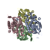
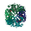
- Assembly
| Deposited unit | 
| ||||||||
|---|---|---|---|---|---|---|---|---|---|
| 1 |
| ||||||||
| 2 | 
| ||||||||
| 3 | 
| ||||||||
| 4 | 
| ||||||||
| Unit cell |
|
-Components
| #1: Protein | Mass: 26141.189 Da / Num. of mol.: 6 Source method: isolated from a genetically manipulated source Source: (gene. exp.) References: UniProt: P32719, Isomerases; Racemases and epimerases; Acting on carbohydrates and derivatives #2: Sugar | ChemComp-S6P /   Type: D-saccharide / Mass: 262.152 Da / Num. of mol.: 6 Type: D-saccharide / Mass: 262.152 Da / Num. of mol.: 6Source method: isolated from a genetically manipulated source Formula: C6H15O9P #3: Chemical | ChemComp-MG /   Mass: 24.305 Da / Num. of mol.: 6 / Source method: obtained synthetically / Formula: Mg Mass: 24.305 Da / Num. of mol.: 6 / Source method: obtained synthetically / Formula: Mg#4: Water | ChemComp-HOH / |  Mass: 18.015 Da / Num. of mol.: 174 / Source method: isolated from a natural source / Formula: H2O Mass: 18.015 Da / Num. of mol.: 174 / Source method: isolated from a natural source / Formula: H2O |
|---|
-Experimental details
-Experiment
| Experiment | Method: X-RAY DIFFRACTION / Number of used crystals: 1 |
|---|
- Sample preparation
| Crystal | Density Matthews: 2.41 Å3/Da / Density % sol: 49 % |
|---|---|
| Crystal grow | Temperature: 293 K / Method: vapor diffusion / pH: 7 Details: 20% PEG 3350, 0.1 M Succinic acid, pH 7.0, VAPOR DIFFUSION, temperature 293.0K |
-Data collection
| Diffraction | Mean temperature: 100 K |
|---|---|
| Diffraction source | Source: SYNCHROTRON / Site: NSLS  / Beamline: X4A / Wavelength: 0.97915 Å / Beamline: X4A / Wavelength: 0.97915 Å |
| Detector | Type: ADSC QUANTUM 4 / Detector: CCD / Date: Sep 3, 2007 |
| Radiation | Monochromator: Si 111 CHANNEL / Protocol: SINGLE WAVELENGTH / Monochromatic (M) / Laue (L): M / Scattering type: x-ray |
| Radiation wavelength | Wavelength: 0.97915 Å / Relative weight: 1 |
| Reflection | Resolution: 2.2→25 Å / Num. all: 74982 / Num. obs: 74982 / % possible obs: 96.5 % / Observed criterion σ(F): 0 / Observed criterion σ(I): 0 / Redundancy: 5.3 % / Biso Wilson estimate: 23.2 Å2 / Rmerge(I) obs: 0.067 |
- Processing
| Software |
| ||||||||||||||||||||||||||||||||||||
|---|---|---|---|---|---|---|---|---|---|---|---|---|---|---|---|---|---|---|---|---|---|---|---|---|---|---|---|---|---|---|---|---|---|---|---|---|---|
| Refinement | Method to determine structure: MOLECULAR REPLACEMENT Starting model: PDB entry 3CT7 Resolution: 2.2→24.96 Å / Rfactor Rfree error: 0.004 / Data cutoff high absF: 1665519.13 / Data cutoff low absF: 0 / Isotropic thermal model: RESTRAINED / Cross valid method: THROUGHOUT / σ(F): 0 / σ(I): 0 / Stereochemistry target values: Engh & Huber
| ||||||||||||||||||||||||||||||||||||
| Solvent computation | Solvent model: FLAT MODEL / Bsol: 29.9587 Å2 / ksol: 0.348849 e/Å3 | ||||||||||||||||||||||||||||||||||||
| Displacement parameters | Biso mean: 39.6 Å2
| ||||||||||||||||||||||||||||||||||||
| Refine analyze |
| ||||||||||||||||||||||||||||||||||||
| Refinement step | Cycle: LAST / Resolution: 2.2→24.96 Å
| ||||||||||||||||||||||||||||||||||||
| Refine LS restraints |
| ||||||||||||||||||||||||||||||||||||
| LS refinement shell | Resolution: 2.2→2.28 Å / Rfactor Rfree error: 0.021 / Total num. of bins used: 10
| ||||||||||||||||||||||||||||||||||||
| Xplor file |
|
