 Movie
Movie Controller

[English] 日本語
 Yorodumi
- PDB-2hbm: Structure of the yeast nuclear exosome component, Rrp6p, reveals ... -
+ Open data

- Basic information
| Entry | Database: PDB / ID: 2hbm | |||||||||
|---|---|---|---|---|---|---|---|---|---|---|
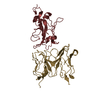
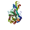
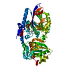
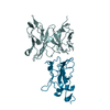
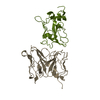
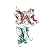
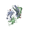
| Title | Structure of the yeast nuclear exosome component, Rrp6p, reveals an interplay between the active site and the HRDC domain; Protein in complex with Mn, Zn, and UMP | |||||||||
 Components Components | Exosome complex exonuclease RRP6 | |||||||||
 Keywords Keywords | HYDROLASE / GENE REGULATION / exosome / RNA metabolism / RNA surveillance / RNA processing | |||||||||
| Function / homology |  Function and homology information Function and homology informationnuclear polyadenylation-dependent antisense transcript catabolic process / nuclear polyadenylation-dependent snoRNA catabolic process / nuclear polyadenylation-dependent snRNA catabolic process / U1 snRNA 3'-end processing / nuclear polyadenylation-dependent mRNA catabolic process / nuclear polyadenylation-dependent CUT catabolic process / U5 snRNA 3'-end processing / TRAMP-dependent tRNA surveillance pathway / exosome (RNase complex) / U4 snRNA 3'-end processing ...nuclear polyadenylation-dependent antisense transcript catabolic process / nuclear polyadenylation-dependent snoRNA catabolic process / nuclear polyadenylation-dependent snRNA catabolic process / U1 snRNA 3'-end processing / nuclear polyadenylation-dependent mRNA catabolic process / nuclear polyadenylation-dependent CUT catabolic process / U5 snRNA 3'-end processing / TRAMP-dependent tRNA surveillance pathway / exosome (RNase complex) / U4 snRNA 3'-end processing / nuclear polyadenylation-dependent rRNA catabolic process / poly(A)-dependent snoRNA 3'-end processing / nuclear exosome (RNase complex) / exonucleolytic trimming to generate mature 3'-end of 5.8S rRNA from tricistronic rRNA transcript (SSU-rRNA, 5.8S rRNA, LSU-rRNA) / histone mRNA catabolic process / nuclear mRNA surveillance / post-transcriptional tethering of RNA polymerase II gene DNA at nuclear periphery / rRNA primary transcript binding / RNA catabolic process / regulation of telomere maintenance / Major pathway of rRNA processing in the nucleolus and cytosol / RNA processing / Hydrolases; Acting on ester bonds; Exoribonucleases producing 5'-phosphomonoesters / 3'-5'-RNA exonuclease activity / single-stranded RNA binding / nucleotide binding / nucleolus / nucleus Similarity search - Function | |||||||||
| Biological species |  | |||||||||
| Method |  X-RAY DIFFRACTION / FOURIER SYNTHESIS / Resolution: 2.7 Å X-RAY DIFFRACTION / FOURIER SYNTHESIS / Resolution: 2.7 Å | |||||||||
 Authors Authors | Midtgaard, S.F. / Assenholt, J. / Jonstrup, A.T. / Van, L.B. / Jensen, T.H. / Brodersen, D.E. | |||||||||
 Citation Citation | Journal: Proc.Natl.Acad.Sci.Usa / Year: 2006 Title: Structure of the nuclear exosome component Rrp6p reveals an interplay between the active site and the HRDC domain. Authors: Midtgaard, S.F. / Assenholt, J. / Jonstrup, A.T. / Van, L.B. / Jensen, T.H. / Brodersen, D.E. | |||||||||
| History |
|
- Structure visualization
| Structure viewer | Molecule: MolmilJmol/JSmol |
|---|
- Downloads & links
-Download
| PDBx/mmCIF format | 2hbm.cif.gz | 100.1 KB | Display | PDBx/mmCIF format |
|---|---|---|---|---|
| PDB format | pdb2hbm.ent.gz | 73.6 KB | Display | PDB format |
| PDBx/mmJSON format | 2hbm.json.gz | Tree view | PDBx/mmJSON format | |
| Others |  Other downloads Other downloads |
-Validation report
| Arichive directory | https://data.pdbj.org/pub/pdb/validation_reports/hb/2hbmftp://data.pdbj.org/pub/pdb/validation_reports/hb/2hbm | HTTPS FTP |
|---|
-Related structure data
| Related structure data | 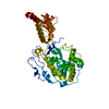 2hbjSC 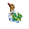 2hbkC 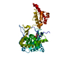 2hblC S: Starting model for refinement C: citing same article ( |
|---|---|
| Similar structure data |


-Links
 PDBj
PDBj


- Assembly
| Deposited unit | 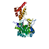
| ||||||||
|---|---|---|---|---|---|---|---|---|---|
| 1 |
| ||||||||
| Unit cell |
| ||||||||
| Details | The biological assembly is the monomer present in the asymmetric unit. |
-Components
| #1: Protein | Mass: 47675.125 Da / Num. of mol.: 1 / Fragment: Rrp6p central fragment, residues 129-536 / Mutation: Y361A Source method: isolated from a genetically manipulated source Source: (gene. exp.) Gene: RRP6 / Plasmid: pET30 Ek/LIC / Production host:  References: UniProt: Q12149, Hydrolases; Acting on ester bonds; Exoribonucleases producing 5'-phosphomonoesters |
|---|---|
| #2: Chemical | ChemComp-ZN /   Mass: 65.409 Da / Num. of mol.: 1 / Source method: obtained synthetically / Formula: Zn Mass: 65.409 Da / Num. of mol.: 1 / Source method: obtained synthetically / Formula: Zn |
| #3: Chemical | ChemComp-MN /   Mass: 54.938 Da / Num. of mol.: 1 / Source method: obtained synthetically / Formula: Mn Mass: 54.938 Da / Num. of mol.: 1 / Source method: obtained synthetically / Formula: Mn |
| #4: Chemical | ChemComp-U5P /   Mass: 324.181 Da / Num. of mol.: 1 / Source method: obtained synthetically / Formula: C9H13N2O9P Mass: 324.181 Da / Num. of mol.: 1 / Source method: obtained synthetically / Formula: C9H13N2O9P |
| #5: Water | ChemComp-HOH /  Mass: 18.015 Da / Num. of mol.: 135 / Source method: isolated from a natural source / Formula: H2O Mass: 18.015 Da / Num. of mol.: 135 / Source method: isolated from a natural source / Formula: H2O |
-Experimental details
-Experiment
| Experiment | Method: X-RAY DIFFRACTION / Number of used crystals: 1 |
|---|
- Sample preparation
| Crystal | Density Matthews: 2.93 Å3/Da / Density % sol: 58.09 % |
|---|---|
| Crystal grow | Temperature: 277 K / Method: vapor diffusion, sitting drop / pH: 7 Details: 12-14% PEG 20000, 0.1M Mes or Hepes, pH 7.0, VAPOR DIFFUSION, SITTING DROP, temperature 277K |
-Data collection
| Diffraction | Mean temperature: 100 K |
|---|---|
| Diffraction source | Source: ROTATING ANODE / Type: ENRAF-NONIUS FR591 / Wavelength: 1.54179 Å |
| Detector | Type: MAR scanner 345 mm plate / Detector: IMAGE PLATE / Date: Jan 9, 2006 / Details: Osmic mirrors |
| Radiation | Monochromator: Osmic mirrors / Protocol: SINGLE WAVELENGTH / Monochromatic (M) / Laue (L): M / Scattering type: x-ray |
| Radiation wavelength | Wavelength: 1.54179 Å / Relative weight: 1 |
| Reflection | Resolution: 2.7→29.61 Å / Num. all: 15522 / Num. obs: 15522 / % possible obs: 98.4 % / Observed criterion σ(F): 0 / Observed criterion σ(I): 0 / Redundancy: 4.1 % / Biso Wilson estimate: 71.43 Å2 / Rmerge(I) obs: 0.061 / Χ2: 0.952 / Net I/σ(I): 14.9 |
| Reflection shell | Resolution: 2.7→2.8 Å / % possible obs: 92.9 % / Redundancy: 3 % / Rmerge(I) obs: 0.666 / Mean I/σ(I) obs: 1.5 / Num. unique all: 1416 / Num. unique obs: 1416 / Χ2: 0.944 / % possible all: 92.9 |
- Processing
| Software |
| ||||||||||||||||||||||||||||||||||||||||||||||||||||||||||||||||||||||||||||||||||||||||||||||||||||||||||||||||||||||||||||||||||||||||||||||||||||||||||||||||||||||||||||||||||||
|---|---|---|---|---|---|---|---|---|---|---|---|---|---|---|---|---|---|---|---|---|---|---|---|---|---|---|---|---|---|---|---|---|---|---|---|---|---|---|---|---|---|---|---|---|---|---|---|---|---|---|---|---|---|---|---|---|---|---|---|---|---|---|---|---|---|---|---|---|---|---|---|---|---|---|---|---|---|---|---|---|---|---|---|---|---|---|---|---|---|---|---|---|---|---|---|---|---|---|---|---|---|---|---|---|---|---|---|---|---|---|---|---|---|---|---|---|---|---|---|---|---|---|---|---|---|---|---|---|---|---|---|---|---|---|---|---|---|---|---|---|---|---|---|---|---|---|---|---|---|---|---|---|---|---|---|---|---|---|---|---|---|---|---|---|---|---|---|---|---|---|---|---|---|---|---|---|---|---|---|---|---|
| Refinement | Method to determine structure: FOURIER SYNTHESIS Starting model: PDB ENTRY 2HBJ Resolution: 2.7→29.61 Å / FOM work R set: 0.796 / Isotropic thermal model: ISOTROPIC / Cross valid method: THROUGHOUT / σ(F): 0 / σ(I): 0 / Stereochemistry target values: Engh & Huber
| ||||||||||||||||||||||||||||||||||||||||||||||||||||||||||||||||||||||||||||||||||||||||||||||||||||||||||||||||||||||||||||||||||||||||||||||||||||||||||||||||||||||||||||||||||||
| Solvent computation | Solvent model: FLAT MODEL / Bsol: 34.9 Å2 | ||||||||||||||||||||||||||||||||||||||||||||||||||||||||||||||||||||||||||||||||||||||||||||||||||||||||||||||||||||||||||||||||||||||||||||||||||||||||||||||||||||||||||||||||||||
| Displacement parameters | Biso mean: 59.7 Å2
| ||||||||||||||||||||||||||||||||||||||||||||||||||||||||||||||||||||||||||||||||||||||||||||||||||||||||||||||||||||||||||||||||||||||||||||||||||||||||||||||||||||||||||||||||||||
| Refine analyze |
| ||||||||||||||||||||||||||||||||||||||||||||||||||||||||||||||||||||||||||||||||||||||||||||||||||||||||||||||||||||||||||||||||||||||||||||||||||||||||||||||||||||||||||||||||||||
| Refinement step | Cycle: LAST / Resolution: 2.7→29.61 Å
| ||||||||||||||||||||||||||||||||||||||||||||||||||||||||||||||||||||||||||||||||||||||||||||||||||||||||||||||||||||||||||||||||||||||||||||||||||||||||||||||||||||||||||||||||||||
| Refine LS restraints |
| ||||||||||||||||||||||||||||||||||||||||||||||||||||||||||||||||||||||||||||||||||||||||||||||||||||||||||||||||||||||||||||||||||||||||||||||||||||||||||||||||||||||||||||||||||||
| LS refinement shell | Refine-ID: X-RAY DIFFRACTION / Total num. of bins used: 29
| ||||||||||||||||||||||||||||||||||||||||||||||||||||||||||||||||||||||||||||||||||||||||||||||||||||||||||||||||||||||||||||||||||||||||||||||||||||||||||||||||||||||||||||||||||||
| Xplor file |
|
