 Movie
Movie Controller

[English] 日本語
 Yorodumi
- PDB-6hbc: Structure of the repeat unit in the network formed by CcmM and Ru... -
+ Open data

- Basic information
| Entry | Database: PDB / ID: 6hbc | ||||||
|---|---|---|---|---|---|---|---|
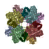
| Title | Structure of the repeat unit in the network formed by CcmM and Rubisco from Synechococcus elongatus | ||||||
 Components Components |
| ||||||
 Keywords Keywords | PROTEIN BINDING / CcmM / M58 / M35 / SSUL domain / Rubisco / Carboxysome | ||||||
| Function / homology |  Function and homology information Function and homology informationstructural constituent of carboxysome shell / photorespiration / carboxysome / ribulose-bisphosphate carboxylase / ribulose-bisphosphate carboxylase activity / reductive pentose-phosphate cycle / carbon fixation / photosynthesis / monooxygenase activity / magnesium ion binding Similarity search - Function | ||||||
| Biological species |  Synechococcus elongatus (bacteria) Synechococcus elongatus (bacteria) | ||||||
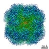
| Method | ELECTRON MICROSCOPY / single particle reconstruction / cryo EM / Resolution: 2.78 Å | ||||||
 Authors Authors | Wang, H. / Yan, X. / Aigner, H. / Bracher, A. / Nguyen, N.D. / Hee, W.Y. / Long, B.M. / Price, G.D. / Hartl, F.U. / Hayer-Hartl, M. | ||||||
 Citation Citation | Journal: Nature / Year: 2019 Title: Rubisco condensate formation by CcmM in β-carboxysome biogenesis. Authors: H Wang / X Yan / H Aigner / A Bracher / N D Nguyen / W Y Hee / B M Long / G D Price / F U Hartl / M Hayer-Hartl /   Abstract: Cells use compartmentalization of enzymes as a strategy to regulate metabolic pathways and increase their efficiency. The α- and β-carboxysomes of cyanobacteria contain ribulose-1,5-bisphosphate ...Cells use compartmentalization of enzymes as a strategy to regulate metabolic pathways and increase their efficiency. The α- and β-carboxysomes of cyanobacteria contain ribulose-1,5-bisphosphate carboxylase/oxygenase (Rubisco)-a complex of eight large (RbcL) and eight small (RbcS) subunits-and carbonic anhydrase. As HCO can diffuse through the proteinaceous carboxysome shell but CO cannot, carbonic anhydrase generates high concentrations of CO for carbon fixation by Rubisco. The shell also prevents access to reducing agents, generating an oxidizing environment. The formation of β-carboxysomes involves the aggregation of Rubisco by the protein CcmM, which exists in two forms: full-length CcmM (M58 in Synechococcus elongatus PCC7942), which contains a carbonic anhydrase-like domain followed by three Rubisco small subunit-like (SSUL) modules connected by flexible linkers; and M35, which lacks the carbonic anhydrase-like domain. It has long been speculated that the SSUL modules interact with Rubisco by replacing RbcS. Here we have reconstituted the Rubisco-CcmM complex and solved its structure. Contrary to expectation, the SSUL modules do not replace RbcS, but bind close to the equatorial region of Rubisco between RbcL dimers, linking Rubisco molecules and inducing phase separation into a liquid-like matrix. Disulfide bond formation in SSUL increases the network flexibility and is required for carboxysome function in vivo. Notably, the formation of the liquid-like condensate of Rubisco is mediated by dynamic interactions with the SSUL domains, rather than by low-complexity sequences, which typically mediate liquid-liquid phase separation in eukaryotes. Indeed, within the pyrenoids of eukaryotic algae, the functional homologues of carboxysomes, Rubisco adopts a liquid-like state by interacting with the intrinsically disordered protein EPYC1. Understanding carboxysome biogenesis will be important for efforts to engineer CO-concentrating mechanisms in plants. | ||||||
| History |
|
- Structure visualization
| Movie |
Movie viewer |
|---|---|
| Structure viewer | Molecule: MolmilJmol/JSmol |
- Downloads & links
-Download
| PDBx/mmCIF format | 6hbc.cif.gz | 237.3 KB | Display | PDBx/mmCIF format |
|---|---|---|---|---|
| PDB format | pdb6hbc.ent.gz | 189.8 KB | Display | PDB format |
| PDBx/mmJSON format | 6hbc.json.gz | Tree view | PDBx/mmJSON format | |
| Others |  Other downloads Other downloads |
-Validation report
| Arichive directory | https://data.pdbj.org/pub/pdb/validation_reports/hb/6hbcftp://data.pdbj.org/pub/pdb/validation_reports/hb/6hbc | HTTPS FTP |
|---|
-Related structure data
| Related structure data | 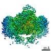 0180MC  6hbaC  6hbbC C: citing same article ( M: map data used to model this data |
|---|---|
| Similar structure data |


-Links
 PDBj
PDBj


- Assembly
| Deposited unit | 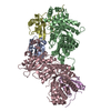
|
|---|---|
| 1 | 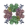
|
-Components
| #1: Protein | Mass: 10550.686 Da / Num. of mol.: 1 / Fragment: SSUL domain 1 Source method: isolated from a genetically manipulated source Source: (gene. exp.) Synechococcus elongatus (strain PCC 7942) (bacteria)Gene: ccmM, Synpcc7942_1423 / Plasmid: pHue / Production host: | ||
|---|---|---|---|
| #2: Protein | Mass: 52516.605 Da / Num. of mol.: 2 / Fragment: Rubisco large subunit Source method: isolated from a genetically manipulated source Source: (gene. exp.) Synechococcus elongatus (strain PCC 7942) (bacteria)Gene: cbbL, rbcL, Synpcc7942_1426 / Plasmid: pET11a / Production host: References: UniProt: Q31NB3, ribulose-bisphosphate carboxylase #3: Protein | Mass: 13349.196 Da / Num. of mol.: 2 / Fragment: Rubisco small subunit Source method: isolated from a genetically manipulated source Source: (gene. exp.) Synechococcus elongatus (strain PCC 7942) (bacteria)Gene: Synpcc7942_1427 / Production host: References: UniProt: Q31NB2, ribulose-bisphosphate carboxylase |
-Experimental details
-Experiment
| Experiment | Method: ELECTRON MICROSCOPY |
|---|---|
| EM experiment | Aggregation state: PARTICLE / 3D reconstruction method: single particle reconstruction |
- Sample preparation
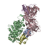
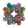
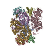
| Component | Name: Repeat unit in the CcmM-Rubisco network consisting of a SSUL domain from CcmM and each two RbcL and two RbcS chains from Rubisco Type: COMPLEX Details: CcmM contains a succession of three highly similar SSUL domains (residues 225-313, 340-428 and 455-539, respectively), which bind to cleft on the surface of Rubisco. Rubisco is a hexadecamer ...Details: CcmM contains a succession of three highly similar SSUL domains (residues 225-313, 340-428 and 455-539, respectively), which bind to cleft on the surface of Rubisco. Rubisco is a hexadecamer of eight RbcL and eight RbcS subunits. The complex has D4 symmetry. The SSUL-RbcL2-RbcS2 repeat units can have one of two orientations (up or down). Thus Rubisco complexes saturated with SSUL domains can have four different configurations (uuuu, uuud, uudd, udud). In reality, some SSUL binding sites are probably left unoccupied. The network is formed by flexible linkers connecting the SSUL domains in CcmM, which then interlink Rubisco hexadecamers. Entity ID: all / Source: RECOMBINANT | |||||||||||||||||||||||||
|---|---|---|---|---|---|---|---|---|---|---|---|---|---|---|---|---|---|---|---|---|---|---|---|---|---|---|
| Molecular weight | Experimental value: NO | |||||||||||||||||||||||||
| Source (natural) | Organism: Synechococcus elongatus PCC 7942 (bacteria) / Organelle: Carboxysome | |||||||||||||||||||||||||
| Source (recombinant) | Organism: | |||||||||||||||||||||||||
| Buffer solution | pH: 8 / Details: Solutions were made fresh | |||||||||||||||||||||||||
| Buffer component |
| |||||||||||||||||||||||||
| Specimen | Conc.: 5 mg/ml / Embedding applied: NO / Shadowing applied: NO / Staining applied: NO / Vitrification applied: YES Details: This sample contained 10 nm gold beads and was not monodisperse | |||||||||||||||||||||||||
| Specimen support | Grid material: COPPER / Grid mesh size: 300 divisions/in. / Grid type: Quantifoil R2/1 | |||||||||||||||||||||||||
| Vitrification | Instrument: FEI VITROBOT MARK IV / Cryogen name: ETHANE / Humidity: 90 % / Chamber temperature: 298 K / Details: blot for 3 seconds before plunging |
- Electron microscopy imaging
| Experimental equipment |  Model: Titan Krios / Image courtesy: FEI Company |
|---|---|
| Microscopy | Model: FEI TITAN KRIOS |
| Electron gun | Electron source:  FIELD EMISSION GUN / Accelerating voltage: 300 kV / Illumination mode: FLOOD BEAM FIELD EMISSION GUN / Accelerating voltage: 300 kV / Illumination mode: FLOOD BEAM |
| Electron lens | Mode: BRIGHT FIELD |
| Image recording | Average exposure time: 0.15 sec. / Electron dose: 1.05 e/Å2 / Film or detector model: GATAN K2 QUANTUM (4k x 4k) |
- Processing
| EM software |
| ||||||||||||
|---|---|---|---|---|---|---|---|---|---|---|---|---|---|
| CTF correction | Type: PHASE FLIPPING AND AMPLITUDE CORRECTION | ||||||||||||
| Symmetry | Point symmetry: C1 (asymmetric) | ||||||||||||
| 3D reconstruction | Resolution: 2.78 Å / Resolution method: FSC 0.143 CUT-OFF / Num. of particles: 78916 / Symmetry type: POINT | ||||||||||||
| Atomic model building | Protocol: AB INITIO MODEL / Space: RECIPROCAL / Target criteria: Average Fourier shell correlation Details: The cryoEM density for the repeat unit was masked by Refmac to the coordinates and converted into structure factors by Refmac. The model was adjusted with coot. This model was submitted to ...Details: The cryoEM density for the repeat unit was masked by Refmac to the coordinates and converted into structure factors by Refmac. The model was adjusted with coot. This model was submitted to restrained refinement with Refmac against the structure factors. |
