 Movie
Movie Controller

[English] 日本語
 Yorodumi
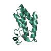
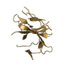
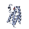
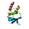
- PDB-6hbb: Crystal Structure of the small subunit-like domain 1 of CcmM from... -
+ Open data

- Basic information
| Entry | Database: PDB / ID: 6hbb | ||||||
|---|---|---|---|---|---|---|---|
| Title | Crystal Structure of the small subunit-like domain 1 of CcmM from Synechococcus elongatus (strain PCC 7942) | ||||||
 Components Components | Carbon dioxide concentrating mechanism protein CcmM | ||||||
 Keywords Keywords | PROTEIN BINDING / alpha-beta structure / Rubisco / Carboxysome | ||||||
| Function / homology |  Function and homology information Function and homology informationstructural constituent of carboxysome shell / carboxysome / carbon fixation / photosynthesis Similarity search - Function | ||||||
| Biological species |  Synechococcus elongatus (bacteria) Synechococcus elongatus (bacteria) | ||||||
| Method |  X-RAY DIFFRACTION / SYNCHROTRON / MOLECULAR REPLACEMENT / molecular replacement / Resolution: 1.2 Å X-RAY DIFFRACTION / SYNCHROTRON / MOLECULAR REPLACEMENT / molecular replacement / Resolution: 1.2 Å | ||||||
 Authors Authors | Wang, H. / Yan, X. / Aigner, H. / Bracher, A. / Nguyen, N.D. / Hee, W.Y. / Long, B.M. / Price, G.D. / Hartl, F.U. / Hayer-Hartl, M. | ||||||
 Citation Citation | Journal: Nature / Year: 2019 Title: Rubisco condensate formation by CcmM in β-carboxysome biogenesis. Authors: H Wang / X Yan / H Aigner / A Bracher / N D Nguyen / W Y Hee / B M Long / G D Price / F U Hartl / M Hayer-Hartl /   Abstract: Cells use compartmentalization of enzymes as a strategy to regulate metabolic pathways and increase their efficiency. The α- and β-carboxysomes of cyanobacteria contain ribulose-1,5-bisphosphate ...Cells use compartmentalization of enzymes as a strategy to regulate metabolic pathways and increase their efficiency. The α- and β-carboxysomes of cyanobacteria contain ribulose-1,5-bisphosphate carboxylase/oxygenase (Rubisco)-a complex of eight large (RbcL) and eight small (RbcS) subunits-and carbonic anhydrase. As HCO can diffuse through the proteinaceous carboxysome shell but CO cannot, carbonic anhydrase generates high concentrations of CO for carbon fixation by Rubisco. The shell also prevents access to reducing agents, generating an oxidizing environment. The formation of β-carboxysomes involves the aggregation of Rubisco by the protein CcmM, which exists in two forms: full-length CcmM (M58 in Synechococcus elongatus PCC7942), which contains a carbonic anhydrase-like domain followed by three Rubisco small subunit-like (SSUL) modules connected by flexible linkers; and M35, which lacks the carbonic anhydrase-like domain. It has long been speculated that the SSUL modules interact with Rubisco by replacing RbcS. Here we have reconstituted the Rubisco-CcmM complex and solved its structure. Contrary to expectation, the SSUL modules do not replace RbcS, but bind close to the equatorial region of Rubisco between RbcL dimers, linking Rubisco molecules and inducing phase separation into a liquid-like matrix. Disulfide bond formation in SSUL increases the network flexibility and is required for carboxysome function in vivo. Notably, the formation of the liquid-like condensate of Rubisco is mediated by dynamic interactions with the SSUL domains, rather than by low-complexity sequences, which typically mediate liquid-liquid phase separation in eukaryotes. Indeed, within the pyrenoids of eukaryotic algae, the functional homologues of carboxysomes, Rubisco adopts a liquid-like state by interacting with the intrinsically disordered protein EPYC1. Understanding carboxysome biogenesis will be important for efforts to engineer CO-concentrating mechanisms in plants. | ||||||
| History |
|
- Structure visualization
| Structure viewer | Molecule: MolmilJmol/JSmol |
|---|
- Downloads & links
-Download
| PDBx/mmCIF format | 6hbb.cif.gz | 94.8 KB | Display | PDBx/mmCIF format |
|---|---|---|---|---|
| PDB format | pdb6hbb.ent.gz | 71.8 KB | Display | PDB format |
| PDBx/mmJSON format | 6hbb.json.gz | Tree view | PDBx/mmJSON format | |
| Others |  Other downloads Other downloads |
-Validation report
| Arichive directory | https://data.pdbj.org/pub/pdb/validation_reports/hb/6hbbftp://data.pdbj.org/pub/pdb/validation_reports/hb/6hbb | HTTPS FTP |
|---|
-Related structure data
| Related structure data | 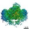 0180C  6hbaSC  6hbcC S: Starting model for refinement C: citing same article ( |
|---|---|
| Similar structure data |


-Links
 PDBj
PDBj


- Assembly
| Deposited unit | 
| ||||||||
|---|---|---|---|---|---|---|---|---|---|
| 1 | 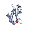
| ||||||||
| 2 | 
| ||||||||
| Unit cell |
|
-Components
| #1: Protein | Mass: 10550.686 Da / Num. of mol.: 2 / Fragment: SSUL domain 1 Source method: isolated from a genetically manipulated source Source: (gene. exp.) Synechococcus elongatus (strain PCC 7942) (bacteria)Strain: PCC 7942 / Gene: ccmM, Synpcc7942_1423 / Plasmid: pHue / Production host: #2: Chemical | ChemComp-SO4 /   Mass: 96.063 Da / Num. of mol.: 7 / Source method: obtained synthetically / Formula: SO4 Mass: 96.063 Da / Num. of mol.: 7 / Source method: obtained synthetically / Formula: SO4#3: Water | ChemComp-HOH / |  Mass: 18.015 Da / Num. of mol.: 162 / Source method: isolated from a natural source / Formula: H2O Mass: 18.015 Da / Num. of mol.: 162 / Source method: isolated from a natural source / Formula: H2O |
|---|
-Experimental details
-Experiment
| Experiment | Method: X-RAY DIFFRACTION / Number of used crystals: 1 |
|---|
- Sample preparation
| Crystal | Density Matthews: 1.98 Å3/Da / Density % sol: 37.77 % / Mosaicity: 0.04 ° |
|---|---|
| Crystal grow | Temperature: 277 K / Method: vapor diffusion / pH: 4.5 Details: 1.95 M ammonium sulfate and 0.1 M Na-acetate pH 4.5 |
-Data collection
| Diffraction | Mean temperature: 100 K | ||||||||||||||||||||||||
|---|---|---|---|---|---|---|---|---|---|---|---|---|---|---|---|---|---|---|---|---|---|---|---|---|---|
| Diffraction source | Source: SYNCHROTRON / Site: ESRF  / Beamline: MASSIF-1 / Wavelength: 0.966 Å / Beamline: MASSIF-1 / Wavelength: 0.966 Å | ||||||||||||||||||||||||
| Detector | Type: DECTRIS PILATUS3 2M / Detector: PIXEL / Date: Jul 21, 2015 | ||||||||||||||||||||||||
| Radiation | Protocol: SINGLE WAVELENGTH / Monochromatic (M) / Laue (L): M / Scattering type: x-ray | ||||||||||||||||||||||||
| Radiation wavelength | Wavelength: 0.966 Å / Relative weight: 1 | ||||||||||||||||||||||||
| Reflection | Resolution: 1.2→43.91 Å / Num. obs: 48992 / % possible obs: 96 % / Redundancy: 2.9 % / CC1/2: 0.997 / Rmerge(I) obs: 0.06 / Rpim(I) all: 0.041 / Rrim(I) all: 0.072 / Net I/σ(I): 9.4 / Num. measured all: 140389 | ||||||||||||||||||||||||
| Reflection shell | Diffraction-ID: 1
|
-Phasing
| Phasing | Method: molecular replacement | ||||||
|---|---|---|---|---|---|---|---|
| Phasing MR |
|
- Processing
| Software |
| |||||||||||||||||||||||||||||||||||||||||||||||||||||||||||||||||||||||||||
|---|---|---|---|---|---|---|---|---|---|---|---|---|---|---|---|---|---|---|---|---|---|---|---|---|---|---|---|---|---|---|---|---|---|---|---|---|---|---|---|---|---|---|---|---|---|---|---|---|---|---|---|---|---|---|---|---|---|---|---|---|---|---|---|---|---|---|---|---|---|---|---|---|---|---|---|---|
| Refinement | Method to determine structure: MOLECULAR REPLACEMENT Starting model: 6HBA Resolution: 1.2→30 Å / Cor.coef. Fo:Fc: 0.968 / Cor.coef. Fo:Fc free: 0.956 / SU B: 1.464 / SU ML: 0.03 / SU R Cruickshank DPI: 0.0451 / Cross valid method: THROUGHOUT / σ(F): 0 / ESU R: 0.045 / ESU R Free: 0.046 Details: HYDROGENS HAVE BEEN ADDED IN THE RIDING POSITIONS U VALUES : REFINED INDIVIDUALLY
| |||||||||||||||||||||||||||||||||||||||||||||||||||||||||||||||||||||||||||
| Solvent computation | Ion probe radii: 0.8 Å / Shrinkage radii: 0.8 Å / VDW probe radii: 1.2 Å | |||||||||||||||||||||||||||||||||||||||||||||||||||||||||||||||||||||||||||
| Displacement parameters | Biso max: 74.01 Å2 / Biso mean: 15.226 Å2 / Biso min: 5.18 Å2
| |||||||||||||||||||||||||||||||||||||||||||||||||||||||||||||||||||||||||||
| Refinement step | Cycle: final / Resolution: 1.2→30 Å
| |||||||||||||||||||||||||||||||||||||||||||||||||||||||||||||||||||||||||||
| Refine LS restraints |
| |||||||||||||||||||||||||||||||||||||||||||||||||||||||||||||||||||||||||||
| LS refinement shell | Resolution: 1.2→1.232 Å / Rfactor Rfree error: 0 / Total num. of bins used: 20
|
