 Movie
Movie Controller

[English] 日本語
 Yorodumi
- PDB-6gbv: A fast recovering full-length LOV protein (DsLOV) from the marine... -
+ Open data

- Basic information
| Entry | Database: PDB / ID: 6gbv | ||||||
|---|---|---|---|---|---|---|---|
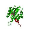
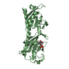
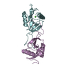
| Title | A fast recovering full-length LOV protein (DsLOV) from the marine phototrophic bacterium Dinoroseobacter shibae (Dark state) - M49T mutant | ||||||
 Components Components | Putative blue-light photoreceptor | ||||||
 Keywords Keywords | SIGNALING PROTEIN / LIGHT-OXYGEN-VOLTAGE / LOV / PAS | ||||||
| Function / homology |  Function and homology information Function and homology information | ||||||
| Biological species |  Dinoroseobacter shibae (bacteria) Dinoroseobacter shibae (bacteria) | ||||||
| Method |  X-RAY DIFFRACTION / SYNCHROTRON / MOLECULAR REPLACEMENT / Resolution: 1.63 Å X-RAY DIFFRACTION / SYNCHROTRON / MOLECULAR REPLACEMENT / Resolution: 1.63 Å | ||||||
 Authors Authors | Granzin, J. / Batra-Safferling, R. / Roellen, K. | ||||||
 Citation Citation | Journal: Biochemistry / Year: 2018 Title: Mechanistic Basis of the Fast Dark Recovery of the Short LOV Protein DsLOV from Dinoroseobacter shibae. Authors: Fettweiss, T. / Rollen, K. / Granzin, J. / Reiners, O. / Endres, S. / Drepper, T. / Willbold, D. / Jaeger, K.E. / Batra-Safferling, R. / Krauss, U. | ||||||
| History |
|
- Structure visualization
| Structure viewer | Molecule: MolmilJmol/JSmol |
|---|
- Downloads & links
-Download
| PDBx/mmCIF format | 6gbv.cif.gz | 65.6 KB | Display | PDBx/mmCIF format |
|---|---|---|---|---|
| PDB format | pdb6gbv.ent.gz | 46.4 KB | Display | PDB format |
| PDBx/mmJSON format | 6gbv.json.gz | Tree view | PDBx/mmJSON format | |
| Others |  Other downloads Other downloads |
-Validation report
| Arichive directory | https://data.pdbj.org/pub/pdb/validation_reports/gb/6gbvftp://data.pdbj.org/pub/pdb/validation_reports/gb/6gbv | HTTPS FTP |
|---|
-Related structure data
| Related structure data | 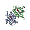 6gayC 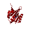 6gb3C 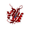 6gbaC 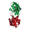 4kukS S: Starting model for refinement C: citing same article ( |
|---|---|
| Similar structure data |


-Links
 PDBj
PDBj

- Assembly
| Deposited unit | 
| ||||||||
|---|---|---|---|---|---|---|---|---|---|
| 1 | 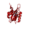
| ||||||||
| Unit cell |
| ||||||||
| Components on special symmetry positions |
|
-Components
| #1: Protein | Mass: 16830.828 Da / Num. of mol.: 1 / Mutation: M49T Source method: isolated from a genetically manipulated source Details: C-terminal hexa-histidine-tagged fusion proteins (tag sequence: LEHHHHHH) in E. coli BL21(DE3) Source: (gene. exp.) Dinoroseobacter shibae (strain DSM 16493 / NCIMB 14021 / DFL 12) (bacteria)Strain: DSM 16493 / NCIMB 14021 / DFL 12 / Gene: Dshi_2006 Production host: References: UniProt: A8LP63 |
|---|---|
| #2: Chemical | ChemComp-FMN /   Mass: 456.344 Da / Num. of mol.: 1 / Source method: obtained synthetically / Formula: C17H21N4O9P Mass: 456.344 Da / Num. of mol.: 1 / Source method: obtained synthetically / Formula: C17H21N4O9P |
| #3: Chemical | ChemComp-PO4 /   Mass: 94.971 Da / Num. of mol.: 1 / Source method: obtained synthetically / Formula: PO4 Mass: 94.971 Da / Num. of mol.: 1 / Source method: obtained synthetically / Formula: PO4 |
| #4: Water | ChemComp-HOH /  Mass: 18.015 Da / Num. of mol.: 78 / Source method: isolated from a natural source / Formula: H2O Mass: 18.015 Da / Num. of mol.: 78 / Source method: isolated from a natural source / Formula: H2O |
-Experimental details
-Experiment
| Experiment | Method: X-RAY DIFFRACTION / Number of used crystals: 1 |
|---|
- Sample preparation
| Crystal | Density Matthews: 1.9 Å3/Da / Density % sol: 35.31 % |
|---|---|
| Crystal grow | Temperature: 292.15 K / Method: vapor diffusion, sitting drop / pH: 7.5 Details: 0.1 M HEPES sodium, 0.8 M Sodium phosphate monobasic monohydrate, 0.8 M Potassium phosphate monobasic |
-Data collection
| Diffraction | Mean temperature: 100 K |
|---|---|
| Diffraction source | Source: SYNCHROTRON / Site: ESRF  / Beamline: MASSIF-3 / Wavelength: 0.9677 Å / Beamline: MASSIF-3 / Wavelength: 0.9677 Å |
| Detector | Type: DECTRIS EIGER X 4M / Detector: PIXEL / Date: Feb 17, 2017 |
| Radiation | Protocol: SINGLE WAVELENGTH / Monochromatic (M) / Laue (L): M / Scattering type: x-ray |
| Radiation wavelength | Wavelength: 0.9677 Å / Relative weight: 1 |
| Reflection | Resolution: 1.63→45.52 Å / Num. obs: 15724 / % possible obs: 98.4 % / Redundancy: 4.7 % / Biso Wilson estimate: 16.7 Å2 / CC1/2: 0.999 / Rmerge(I) obs: 0.064 / Rrim(I) all: 0.072 / Net I/σ(I): 13.2 |
| Reflection shell | Resolution: 1.63→1.66 Å / Redundancy: 4.8 % / Rmerge(I) obs: 0.708 / Mean I/σ(I) obs: 2.1 / Num. unique obs: 760 / CC1/2: 0.699 / Rrim(I) all: 0.794 / % possible all: 97.1 |
- Processing
| Software |
| |||||||||||||||||||||||||||||||||||||||||||||||||
|---|---|---|---|---|---|---|---|---|---|---|---|---|---|---|---|---|---|---|---|---|---|---|---|---|---|---|---|---|---|---|---|---|---|---|---|---|---|---|---|---|---|---|---|---|---|---|---|---|---|---|
| Refinement | Method to determine structure: MOLECULAR REPLACEMENT Starting model: 4KUK Resolution: 1.63→26.2 Å / SU ML: 0.16 / Cross valid method: FREE R-VALUE / σ(F): 1.37 / Phase error: 20.53
| |||||||||||||||||||||||||||||||||||||||||||||||||
| Solvent computation | Shrinkage radii: 0.9 Å / VDW probe radii: 1.2 Å | |||||||||||||||||||||||||||||||||||||||||||||||||
| Refinement step | Cycle: LAST / Resolution: 1.63→26.2 Å
| |||||||||||||||||||||||||||||||||||||||||||||||||
| Refine LS restraints |
| |||||||||||||||||||||||||||||||||||||||||||||||||
| LS refinement shell |
|
